Skip to Content
Merck
All Photos(2)

Key Documents

HPA011025

Sigma-Aldrich

Anti-COL18A1 antibody produced in rabbit

Prestige Antibodies® Powered by Atlas Antibodies, affinity isolated antibody, buffered aqueous glycerol solution

Synonym(s):

Anti-Collagen α-1(XVIII) chain precursor antibody produced in rabbit

Sign Into View Organizational & Contract Pricing


About This Item

UNSPSC Code:
12352203
Human Protein Atlas Number:

biological source

rabbit

conjugate

unconjugated

antibody form

affinity isolated antibody

antibody product type

primary antibodies

clone

polyclonal

product line

Prestige Antibodies® Powered by Atlas Antibodies

form

buffered aqueous glycerol solution

species reactivity

human

technique(s)

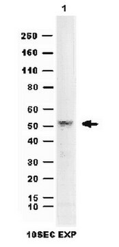
immunoblotting: 0.04-0.4 μg/mL
immunohistochemistry: 1:50-1:200

immunogen sequence

KDELLFPSWEALFSGSEGPLKPGARIFSFDGKDVLRHPTWPQKSVWHGSDPNGRRLTESYCETWRTEAPSATGQASSLLGGRLLGQSAASCHHAYIVLCIENSFM

UniProt accession no.

shipped in

wet ice

storage temp.

−20°C

Gene Information

human ... COL18A1(80781)

General description

COL18A1 (collagen, type XVIII, α 1) codes for the α chain of collagen XVIII. It belongs to multiplexins (multiple triple-helix domains with interruptions) subgroup of collagen family. It is a proteoglycan collagen, which is non-fibrillar in nature. Its N- and C- termini are non-collagenous in nature and are intervened by alternating 10 collagenous and 9 non-collagenous repeats. The N-terminal contains thrombospondin-1-like motif, and a motif of 120 amino acids called frizzled motif, which has a cysteine-rich domain (CRD). This gene is located on human chromosome 21q22.3, and has 43 exons. Due to alternative splicing, this protein has three isoforms named, NC11-303, NC11-493 and NC11-728, which differ partly at their N-terminal and signal peptide. NC11-303 is expressed in fetal brain, kidney and retina; NC11-493 is predominantly expressed in adult and fetal liver, and NC11-728 is widely expressed, though predominant expression being in liver and lung.

Immunogen

Collagen α-1(XVIII) chain precursor recombinant protein epitope signature tag (PrEST)

Application

All Prestige Antibodies Powered by Atlas Antibodies are developed and validated by the Human Protein Atlas (HPA) project and as a result, are supported by the most extensive characterization in the industry.

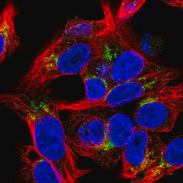
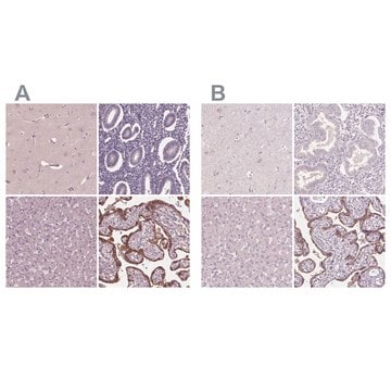
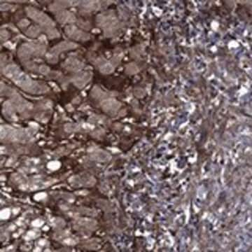
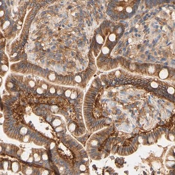
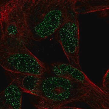
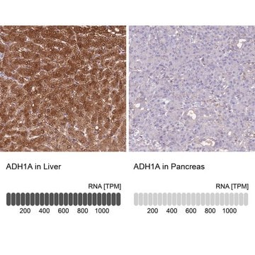
The Human Protein Atlas project can be subdivided into three efforts: Human Tissue Atlas, Cancer Atlas, and Human Cell Atlas. The antibodies that have been generated in support of the Tissue and Cancer Atlas projects have been tested by immunohistochemistry against hundreds of normal and disease tissues and through the recent efforts of the Human Cell Atlas project, many have been characterized by immunofluorescence to map the human proteome not only at the tissue level but now at the subcellular level. These images and the collection of this vast data set can be viewed on the Human Protein Atlas (HPA) site by clicking on the Image Gallery link. We also provide Prestige Antibodies® protocols and other useful information.

Biochem/physiol Actions

COL18A1 (collagen, type XVIII, α 1) is responsible for defining the structure of retina and regulates the closure of neural tube. During embryogenesis, it is essential for normal eye development, and is expressed in the basement membrane of iris, vitreous and retina. It is also a part of the basement membrane of kidney, and is involved in teeth development. It is also involved in the migration of neurons. COL18A1 plays a role in normal development of brain, and mutations in this gene lead to structural abnormalities in brain. This protein shows marked increase in the cerebrospinal fluid (CSF) of patients with severe traumatic brain injury, and thus, COL18A1 might be involved in the response of central nervous system to any injury. Mutations in this gene lead to Knobloch Syndrome (KS), which is a rare autosomal recessive disorder.

Features and Benefits

Prestige Antibodies® are highly characterized and extensively validated antibodies with the added benefit of all available characterization data for each target being accessible via the Human Protein Atlas portal linked just below the product name at the top of this page. The uniqueness and low cross-reactivity of the Prestige Antibodies® to other proteins are due to a thorough selection of antigen regions, affinity purification, and stringent selection. Prestige antigen controls are available for every corresponding Prestige Antibody and can be found in the linkage section.

Every Prestige Antibody is tested in the following ways:
  • IHC tissue array of 44 normal human tissues and 20 of the most common cancer type tissues.
  • Protein array of 364 human recombinant protein fragments.

Linkage

Corresponding Antigen APREST72542.

Physical form

Solution in phosphate-buffered saline, pH 7.2, containing 40% glycerol and 0.02% sodium azide

Legal Information

Prestige Antibodies is a registered trademark of Merck KGaA, Darmstadt, Germany

Disclaimer

Unless otherwise stated in our catalog or other company documentation accompanying the product(s), our products are intended for research use only and are not to be used for any other purpose, which includes but is not limited to, unauthorized commercial uses, in vitro diagnostic uses, ex vivo or in vivo therapeutic uses or any type of consumption or application to humans or animals.

Not finding the right product?  

Try our Product Selector Tool.

Storage Class Code

10 - Combustible liquids

WGK

WGK 1

Flash Point(F)

Not applicable

Flash Point(C)

Not applicable

Personal Protective Equipment

dust mask type N95 (US), Eyeshields, Gloves

Choose from one of the most recent versions:

Certificates of Analysis (COA)

Lot/Batch Number

Don't see the Right Version?

If you require a particular version, you can look up a specific certificate by the Lot or Batch number.

Already Own This Product?

Find documentation for the products that you have recently purchased in the Document Library.

Visit the Document Library

Alireza Haghighi et al.
PloS one, 9(11), e112747-e112747 (2014-11-14)
The aim of this study was to identify the genetic basis of a chorioretinal dystrophy with high myopia of unknown origin in a child of a consanguineous marriage. The proband and ten family members of Iranian ancestry participated in this
Hao Chen et al.
BioMed research international, 2013, 402375-402375 (2013-10-04)
Recent studies have suggested that endogenous angiogenesis inhibitor endostatin/collagen XVIII might play an important role in the secondary brain injury following traumatic brain injury (TBI). In this study, we measured endostatin/collagen XVIII concentrations serially for 1 week after hospitalization by
P M Hägg et al.
The Journal of biological chemistry, 273(28), 17824-17831 (1998-07-04)
The human gene for the alpha1 chain of type XV collagen (COL15A1) is about 145 kilobases in size and contains 42 exons. The promoter is characterized by the lack of a TATAA motif and the presence of several Sp1 binding
Maria Rita Passos-Bueno et al.
Anais da Academia Brasileira de Ciencias, 78(1), 123-131 (2006-03-15)
Collagen XVIII, a proteoglycan, is a component of basement membranes (BMs). There are three distinct isoforms that differ only by their N-terminal, but with a specific pattern of tissue and developmental expression. Cleavage of its C-terminal produces endostatin, an inhibitor
Ahmet Okay Caglayan et al.
Pediatric neurology, 51(6), 806-813 (2014-12-03)
Knobloch syndrome is a rare, autosomal recessive, developmental disorder characterized by stereotyped ocular abnormalities with or without occipital skull deformities (encephalocele, bone defects, and cutis aplasia). Although there is clear heterogeneity in clinical presentation, central nervous system malformations, aside from

Questions

Reviews

No rating value

Active Filters

Our team of scientists has experience in all areas of research including Life Science, Material Science, Chemical Synthesis, Chromatography, Analytical and many others.

Contact Technical Service