Skip to Content
Merck
All Photos(1)

Documents

18772

Sigma-Aldrich

Anti-Rabbit IgG - Atto 488 antibody produced in goat

whole molecule, 1 mg/mL protein

Synonym(s):

Atto 488-[goat-Anti-rabbit IgG], whole molecule

Sign Into View Organizational & Contract Pricing


About This Item

UNSPSC Code:
12352203
NACRES:
NA.46

conjugate

Atto 488 conjugate

antibody form

affinity isolated antibody

antibody product type

secondary antibodies

clone

polyclonal

contains

50% glycerol

species reactivity

rabbit

concentration

1 mg/mL protein

technique(s)

immunocytochemistry: 1:500
immunofluorescence: 1:200
immunohistochemistry: suitable

fluorescence

λex 485 nm; λem 552 nm in PBS

shipped in

wet ice

storage temp.

−20°C

target post-translational modification

unmodified

General description

Goat polyclonal anti-Rabbit IgG (whole molecule)-Atto 488 antibody antibody reats with rabbit IgG in vitro, in rabbit serum and biological fluids.
Rabbit IgG is a plasma B cell derived antibody isotype defined by its heavy chain. IgG is the most abundant antibody isotype found in rabbit serum. IgG crosses the placental barrier, is a complement activator and binds to the Fc-receptors on phagocytic cells. The level of IgG may vary with the status of disease or infection.
Atto 488 is a labeling dye with high molecular absorption (90,000) and quantum yield (0.80) as well as sufficient Stokes shift between excitation and emission maximum. It is optimized for excitation with an argon laser, and is characterized by high photostability. Atto fluorescent labels are designed for high sensitivity applications, including single molecule detection. Atto labels have rigid structures that do not show any cis-trans-isomerization. Thus these labels display exceptional intensity with minimal spectral shift on conjugation.

Immunogen

Rabbit IgG

Application

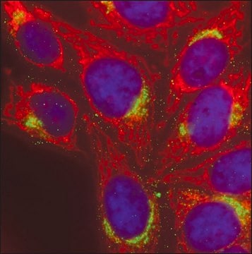
Anti-Rabbit IgG has been used for immunocytochemistry applications at a dilution of 1:500 in neural precursor cells.
Goat polyclonal anti-Rabbit IgG (whole molecule)-Atto 488 antibody may be used to detect and quantitate the level of IgG in rabbit serum and biological fluids via fluorogenic immunochemical or immunohistochemical techniques. It may also be used as a secondary antibody in assays that use rabbit IgG as the primary antibody. Atto 488 goat anti-rabbit IgG antibody was used for immunofluorescence to detect L. monocytogenes in mouse tissue sections following incubation with an anti-listeria antibody. Antibody was used at a 1:200 dilution.

Physical form

Atto 488 goat anti-rabbit IgG (whole molecule) is provided in unit sizes of 1 ml as 1 mg/ml solutions in 0.1 M sodium phosphate, 0.1 M NaCl, pH 7.5, containing 5 mM sodium azide as a preservative.

Analysis Note

Unconjugated dye ≤5% of total fluorescence; dye-to-protein ratio >2.

Legal Information

This product is for Research use only. In case of intended commercialization, please contact the IP-holder (ATTO-TEC GmbH, Germany) for licensing.

Disclaimer

Unless otherwise stated in our catalog or other company documentation accompanying the product(s), our products are intended for research use only and are not to be used for any other purpose, which includes but is not limited to, unauthorized commercial uses, in vitro diagnostic uses, ex vivo or in vivo therapeutic uses or any type of consumption or application to humans or animals.

Not finding the right product?  

Try our Product Selector Tool.

Storage Class Code

10 - Combustible liquids

WGK

WGK 3

Flash Point(F)

Not applicable

Flash Point(C)

Not applicable


Certificates of Analysis (COA)

Search for Certificates of Analysis (COA) by entering the products Lot/Batch Number. Lot and Batch Numbers can be found on a product’s label following the words ‘Lot’ or ‘Batch’.

Already Own This Product?

Find documentation for the products that you have recently purchased in the Document Library.

Visit the Document Library

Tatiana D Mayorova et al.
Scientific reports, 11(1), 23343-23343 (2021-12-04)
Placozoa is a phylum of non-bilaterian marine animals. These small, flat organisms adhere to the substrate via their densely ciliated ventral epithelium, which mediates mucociliary locomotion and nutrient uptake. They have only six morphological cell types, including one, fiber cells
Lin Wang et al.
Oncology letters, 23(5), 149-149 (2022-03-31)
Throughout the world, numerous individuals are infected with Toxoplasma gondii, which may improve immunity against cancer. Furthermore, microRNAs (miRs) may be differentially expressed in the host upon infection with T. gondii. In the present study, RNA-sequencing analysis and reverse transcription-quantitative
Javier A Carrero et al.
Infection and immunity, 77(10), 4371-4382 (2009-08-12)
Listeriolysin O (LLO) is an essential virulence factor for the gram-positive bacterium Listeria monocytogenes. Our goal was to determine if altering the topology of LLO would alter the virulence and toxicity of L. monocytogenes in vivo. A recombinant strain was
Janin Lautenschläger et al.
Nature communications, 9(1), 712-712 (2018-02-21)
Alpha-synuclein is known to bind to small unilamellar vesicles (SUVs) via its N terminus, which forms an amphipathic alpha-helix upon membrane interaction. Here we show that calcium binds to the C terminus of alpha-synuclein, therewith increasing its lipid-binding capacity. Using
Jeffrey M Schaub et al.
Langmuir : the ACS journal of surfaces and colloids, 34(49), 14882-14890 (2018-07-26)
Single-stranded DNA (ssDNA) is a critical intermediate in all DNA transactions. Because ssDNA is more flexible than double-stranded (ds) DNA, interactions with ssDNA-binding proteins (SSBs) may significantly compact or elongate the ssDNA molecule. Here, we develop and characterize low-complexity ssDNA

Our team of scientists has experience in all areas of research including Life Science, Material Science, Chemical Synthesis, Chromatography, Analytical and many others.

Contact Technical Service