Skip to Content
Merck
All Photos(6)

Key Documents

HPA019493

Sigma-Aldrich

Anti-LCP1 antibody produced in rabbit

enhanced validation

Prestige Antibodies® Powered by Atlas Antibodies, affinity isolated antibody, buffered aqueous glycerol solution

Synonym(s):

Anti-L-plastin, Anti-LC64P, Anti-LCP-1, Anti-Lymphocyte cytosolic protein 1, Anti-Plastin-2

Sign Into View Organizational & Contract Pricing


About This Item

UNSPSC Code:
12352203
Human Protein Atlas Number:
NACRES:
NA.41

biological source

rabbit

conjugate

unconjugated

antibody form

affinity isolated antibody

antibody product type

primary antibodies

clone

polyclonal

product line

Prestige Antibodies® Powered by Atlas Antibodies

form

buffered aqueous glycerol solution

species reactivity

human, rat

enhanced validation

orthogonal RNAseq
Learn more about Antibody Enhanced Validation

technique(s)

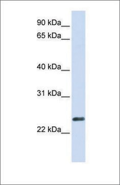
immunoblotting: 0.04-0.4 μg/mL
immunofluorescence: 0.25-2 μg/mL
immunohistochemistry: 1:500-1:1000

immunogen sequence

FAKVDTDGNGYISFNELNDLFKAACLPLPGYRVREITENLMATGDLDQDGRISFDEFIKI

UniProt accession no.

shipped in

wet ice

storage temp.

−20°C

target post-translational modification

unmodified

Gene Information

human ... LCP1(3936)

General description

LCP1 (Lymphocyte cytosolic protein 1) is an actin-binding protein belonging to a large family of actin filament cross-linking proteins. It was firstly identified as an isoform of intestinal brush-border fimbrin. It consists of a calmodulin-like headpiece domain at the N-terminal end, which is equipped with two helix-loop-helix EF-hand Ca2+ binding motifs, followed by two independent actin-binding domains (ABDs) within the same region. It is localized to actin-rich membrane structures.

Immunogen

Plastin-2 recombinant protein epitope signature tag (PrEST)

Application

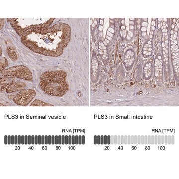
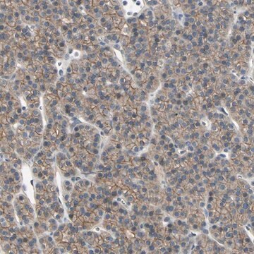
Anti-LCP1 antibody produced in rabbit, a Prestige Antibody, is developed and validated by the Human Protein Atlas (HPA) project . Each antibody is tested by immunohistochemistry against hundreds of normal and disease tissues. These images can be viewed on the Human Protein Atlas (HPA) site by clicking on the Image Gallery link. The antibodies are also tested using immunofluorescence and western blotting. To view these protocols and other useful information about Prestige Antibodies and the HPA, visit sigma.com/prestige.

Biochem/physiol Actions

LCP1 (Lymphocyte cytosolic protein 1) is majorly involved in the actin cytoskeleton assembly through the cross-linking activities. In addition, it is also associated with several activities such as locomotion, adhesion and immune defence, cell-adhesions, and the phagocytosis. It is phosphorylated at the serine residue positions during cytoskeleton organization to control F-actin-binding properties. Expression of LCP1 has been reported in several types of malignant human cells promoting tumor metastasis. It functions as a malignant transformation-associated protein.

Features and Benefits

Prestige Antibodies® are highly characterized and extensively validated antibodies with the added benefit of all available characterization data for each target being accessible via the Human Protein Atlas portal linked just below the product name at the top of this page. The uniqueness and low cross-reactivity of the Prestige Antibodies® to other proteins are due to a thorough selection of antigen regions, affinity purification, and stringent selection. Prestige antigen controls are available for every corresponding Prestige Antibody and can be found in the linkage section.

Every Prestige Antibody is tested in the following ways:
  • IHC tissue array of 44 normal human tissues and 20 of the most common cancer type tissues.
  • Protein array of 364 human recombinant protein fragments.

Linkage

Corresponding Antigen APREST74598

Physical form

Solution in phosphate-buffered saline, pH 7.2, containing 40% glycerol and 0.02% sodium azide

Legal Information

Prestige Antibodies is a registered trademark of Merck KGaA, Darmstadt, Germany

Disclaimer

Unless otherwise stated in our catalog or other company documentation accompanying the product(s), our products are intended for research use only and are not to be used for any other purpose, which includes but is not limited to, unauthorized commercial uses, in vitro diagnostic uses, ex vivo or in vivo therapeutic uses or any type of consumption or application to humans or animals.

Not finding the right product?  

Try our Product Selector Tool.

Storage Class Code

10 - Combustible liquids

WGK

WGK 1

Flash Point(F)

Not applicable

Flash Point(C)

Not applicable


Certificates of Analysis (COA)

Search for Certificates of Analysis (COA) by entering the products Lot/Batch Number. Lot and Batch Numbers can be found on a product’s label following the words ‘Lot’ or ‘Batch’.

Already Own This Product?

Find documentation for the products that you have recently purchased in the Document Library.

Visit the Document Library

Selina M Riplinger et al.
Molecular cancer, 13, 10-10 (2014-01-21)
Tumor cell migration and metastasis require dynamic rearrangements of the actin cytoskeleton. Interestingly, the F-actin cross-linking and stabilizing protein L-plastin, originally described as a leukocyte specific protein, is aberrantly expressed in several non-hematopoietic malignant tumors. Therefore, it has been discussed
Guido H Wabnitz et al.
European journal of immunology, 37(3), 649-662 (2007-02-13)
Rearrangements in the actin cytoskeleton play a pivotal role for costimulation-induced formation of the immunological synapse and T cell activation. Yet, little is known about the actin-binding proteins that link costimulation to rearrangements in the actin cytoskeleton. Here we demonstrate
Bassam Janji et al.
Journal of cell science, 119(Pt 9), 1947-1960 (2006-04-26)
L-plastin, a malignant transformation-associated protein, is a member of a large family of actin filament cross-linkers. Here, we analysed how phosphorylation of L-plastin on Ser5 of the headpiece domain regulates its intracellular distribution and its interaction with F-actin in transfected
Julie Bachmann et al.
PLoS pathogens, 10(4), e1004038-e1004038 (2014-04-20)
Systemic inflammation and sequestration of parasitized erythrocytes are central processes in the pathophysiology of severe Plasmodium falciparum childhood malaria. However, it is still not understood why some children are more at risks to develop malaria complications than others. To identify
Marthe Norreen-Thorsen et al.
Cell reports, 40(2), 111046-111046 (2022-07-14)
The importance of defining cell-type-specific genes is well acknowledged. Technological advances facilitate high-resolution sequencing of single cells, but practical challenges remain. Adipose tissue is composed primarily of adipocytes, large buoyant cells requiring extensive, artefact-generating processing for separation and analysis. Thus

Our team of scientists has experience in all areas of research including Life Science, Material Science, Chemical Synthesis, Chromatography, Analytical and many others.

Contact Technical Service