Skip to Content
Merck
All Photos(2)

Key Documents

MABT538

Sigma-Aldrich

Anti-Lamin A/C Antibody, clone 2E8.2

clone 2E8.2, from mouse

Synonym(s):

Prelamin-A/C, Lamin-A/C, Renal carcinoma antigen NY-REN-32

Sign Into View Organizational & Contract Pricing


About This Item

UNSPSC Code:
12352203
eCl@ss:
32160702

biological source

mouse

Quality Level

antibody form

purified antibody

antibody product type

primary antibodies

clone

2E8.2, monoclonal

species reactivity

human

technique(s)

immunohistochemistry: suitable (paraffin)
western blot: suitable

isotype

IgG1κ

NCBI accession no.

UniProt accession no.

shipped in

ambient

target post-translational modification

unmodified

Gene Information

human ... LMNA(4000)

General description

Lamin-A is a key component of the nuclear lamina, a protein network underlying the inner membrane that determines nuclear shape and size. The same gene (Lmna, Dhe, or Lamn1 in murine species; Gene ID 16905) that encodes lamin-A also codes for the alternatively spliced prelamin-C isoforms (UniProt P48678-2 and P48678-3). Murine lamin-A (UniProt P48678-1) is initially produced as a 665-amino acid prelamin A, which is further processed posttranslationally by four enzymatic steps to yield the mature Lamin-A. Farnesylation of Cys662, endoproteolytic cleavage of the last three amino acids (a.a. 663-665), carboxyl methylation of Cys662, and the final endoproteolytic removal of the remaining propeptide sequence (a.a. 648-662), including the farnesylated Cys662, catalyzed by the ER membrane zinc metalloproteinase ZMPSTE24.

Specificity

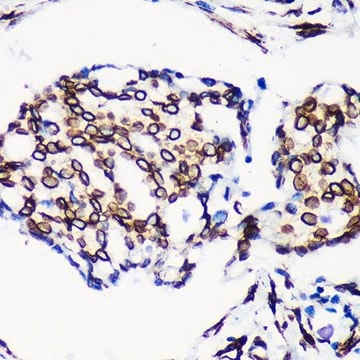
Clone 2E8.2 immunostained nuclear membrane in human tissue sections.

Immunogen

GST-tagged recombinant fragment within the C-terminal half of human Lamin A.

Application

Immunohistochemistry Analysis: A 1:1,000 dilution from a representative lot immunostained nuclear membrane in human cerebellum, kidney, and pancreas tissue sections.
Research Category
Cell Structure
This mouse monoclonal Anti-Lamin A/C Antibody, clone 2E8.2, Cat. No. MABT538 is validated for use in Immunohistochemistry (Paraffin) and Western Blotting for the detection of lamin A/C.

Quality

Evaluated by Western Blotting in A431 cell lysate.

Western Blotting Analysis: 0.5 µg/mL of this antibody detected lamin A/C in 10 µg of A431 cell lysate.

Target description

~70/65 kDa observed. 74.14/73.81/72.22 kDa (perpro-/pre-/mature Lamin A) and 65.14 kDa (Lamin C) calculated. Uncharacterized bands may be observed in some lysate(s).

Linkage

Replaces: MAB3211

Physical form

Format: Purified
Protein G purified.
Purified mouse IgG1κ in buffer containing 0.1 M Tris-Glycine (pH 7.4), 150 mM NaCl with 0.05% sodium azide.

Storage and Stability

Stable for 1 year at 2-8°C from date of receipt.

Other Notes

Concentration: Please refer to lot specific datasheet.

Disclaimer

Unless otherwise stated in our catalog or other company documentation accompanying the product(s), our products are intended for research use only and are not to be used for any other purpose, which includes but is not limited to, unauthorized commercial uses, in vitro diagnostic uses, ex vivo or in vivo therapeutic uses or any type of consumption or application to humans or animals.

Not finding the right product?  

Try our Product Selector Tool.

Storage Class Code

12 - Non Combustible Liquids

WGK

WGK 1


Certificates of Analysis (COA)

Search for Certificates of Analysis (COA) by entering the products Lot/Batch Number. Lot and Batch Numbers can be found on a product’s label following the words ‘Lot’ or ‘Batch’.

Already Own This Product?

Find documentation for the products that you have recently purchased in the Document Library.

Visit the Document Library

Luke W Koblan et al.
Nature, 589(7843), 608-614 (2021-01-08)
Hutchinson-Gilford progeria syndrome (HGPS or progeria) is typically caused by a dominant-negative C•G-to-T•A mutation (c.1824 C>T; p.G608G) in LMNA, the gene that encodes nuclear lamin A. This mutation causes RNA mis-splicing that produces progerin, a toxic protein that induces rapid
Zhuo Zhou et al.
Signal transduction and targeted therapy, 6(1), 382-382 (2021-11-05)
The global coronavirus disease 2019 (COVID-19) pandemic is caused by severe acute respiratory syndrome coronavirus 2 (SARS-CoV-2), a positive-sense RNA virus. How the host immune system senses and responds to SARS-CoV-2 infection remain largely unresolved. Here, we report that SARS-CoV-2

Our team of scientists has experience in all areas of research including Life Science, Material Science, Chemical Synthesis, Chromatography, Analytical and many others.

Contact Technical Service