Proteinase K: Introduction & Applications

What is proteinase K?
Proteinase K is a serine protease that breaks down proteins by hydrolyzing peptide bonds. It is a broad-spectrum protease that can digest a wide variety of proteins, including those that are resistant to other proteases.
Proteinase K is commonly used in molecular biology and biochemistry applications to digest structural proteins and enzymes. It is useful in removing nucleases that can degrade DNA and RNA, as well as in the isolation of intact genomic DNA from various sources. Proteinase K is also used in the analysis of prion protein structure and function (Cronier et al. 2008).
Proteinase K is stable in a wide range of pH, temperature, and can withstand many detergents and denaturing agents. As a result, it is effective for sample preparation in a variety of research and diagnostic applications.
What is proteinase K used for?
Proteinase K is commonly used to isolate DNA or RNA in molecular biology research by degrading unwanted proteins in a sample, such as nucleases, RNases, and DNases. Proteinase K is also used in the process of nucleic acid extraction to break down the protein component of the cell membrane and allow access to the DNA and RNA. It is effective at digesting many types of proteins, including those that are resistant to other types of proteases, such as trypsin.
In addition to molecular biology research, proteinase K is used in a wide range of other applications, including pharmaceutical manufacturing and diagnostic applications. It can be used in the pharmaceutical industry to remove unwanted proteins in their production process. For diagnostics, it is used in the isolation of DNA and RNA from clinical samples for downstream analysis.
Why is proteinase K used in DNA and RNA extraction?
Proteinase K is used in DNA and RNA extraction because it is a proteolytic enzyme that can break down proteins, including those that are present in the cell membrane and nucleus. When using proteinase K for DNA and RNA extraction, it is typically added to a lysis buffer that contains detergents and salts that help to disrupt the cell membrane and release the nucleic acids into the solution. The proteinase K then digests the proteins in the lysed sample, including histones and other chromosomal proteins, which can otherwise interfere with downstream applications such asPCR, RT-PCR, Sanger sequencing, Next Gen Sequencing (NGS), fluorescent in situ hybridization (FISH), and other molecular biology research and diagnostics applications. The use of proteinase K in DNA and RNA extraction helps to ensure the purity and integrity of the sample.
Why is proteinase K used in PCR?
Proteinase K is not typically used directly in a polymerase chain reaction (PCR), but it is used in the DNA extraction step before PCR to improve the quality of the DNA sample.
Proteinase K is effective at breaking down a wide range of proteins, including those that are resistant to other types of proteases, such as trypsin. By removing protein contaminants from the DNA sample before PCR, the efficiency and accuracy of the reaction can be improved, leading to more reliable results. Additionally, the use of proteinase K in DNA extraction can help to reduce the risk of false negatives or other errors in diagnostic testing, where accurate and reliable results are critical.
During PCR, the DNA template is amplified through a series of cycles of denaturation, annealing, and extension. Any impurities in the DNA sample, such as proteins, can interfere with this process by inhibiting the activity of the polymerase enzyme or interfering with the binding of primers to the template.
What is the proteinase K mechanism of action?
Proteinase K is a serine protease that cleaves peptide bonds in proteins. Its mechanism of action involves several steps:
- Binding: Proteinase K first binds to the protein or nucleic acid substrate through non-specific hydrophobic interactions.
- Activation: Once bound, the enzyme undergoes an activation step where a catalytic serine residue is activated by a histidine residue and a water molecule. This leads to the formation of an active site that can cleave peptide bonds.
- Cleavage: The active site of proteinase K cleaves the peptide bond on the carboxylic acid side of the amino acid residues that contain aromatic, aliphatic or hydrophobic amino acid residues. The enzyme can also cleave peptide bonds on the amide side of glycine residues.
- Product release: After cleavage, the products of the reaction (i.e., peptides) are released from the enzyme.
The mechanism of action of proteinase K is similar to other serine proteases, but it is notable for its ability to function under harsh conditions, such as high temperatures and in the presence of detergents.
What is the difference between protease and proteinase K?
Proteases and proteinase K are both proteases that differ in some important ways. Proteases are a class of enzymes that cleave peptide bonds in proteins. There are many different types of proteases, including serine proteases, cysteine proteases, and metalloproteases, among others. These enzymes are involved in many biological processes, including protein degradation, blood coagulation, and digestion. Alternatively, proteinase K is a specific type of serine protease that is widely used to digest proteins and expose the nucleic acids that have been isolated from cells or tissues.
What is the proteinase K DNA extraction method?
The proteinase K DNA extraction method is commonly used to isolate high-quality DNA for downstream applications, such as PCR, sequencing, and cloning. The method is particularly useful for samples that contain high amounts of proteins, such as tissues or cells, as it allows for efficient removal of proteins without damaging the DNA. The proteinase K DNA extraction method typically involves the following steps:
- Sample lysis:Lyse the cells or tissues and release the genomic DNA by adding a lysis buffer to the sample that contains detergents and other reagents that disrupt the cell membrane and break apart protein complexes.
- Protein digestion:Add proteinase K to the sample to digest the proteins that are bound to the DNA. The enzyme is usually added to the sample at a final concentration of 0.2-1 mg/mL and incubated at an optimal temperature (55-65 °C) for 30-60 minutes.
- Deactivation: After the protein digestion is complete, the proteinase K is deactivated by heating the sample to above
95 °C for a few minutes or by adding a protease inhibitor to the sample. - DNA isolation:The DNA can then be isolated from the sample by various methods, such as precipitation with ethanol or isopropanol, or by using a commercial DNA isolation kit
What is the role of proteinase K in covid testing?

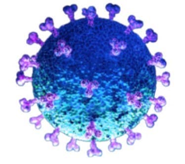
Proteinase K does not directly test for SARS-CoV-2 virus, but it is used in the RNA extraction step upstream in the COVID-19 diagnostic process.
The virus contains genetic material in the form of RNA. To detect the presence of the virus, the test involves first extracting RNA from a patient sample, such as a nasal swab, and then using RT-PCR (reverse transcription polymerase chain reaction) to amplify and detect specific viral RNA sequences. Proteinase K can be used in the RNA extraction process to digest and inactivate RNases, thereby protecting the RNA by eliminating false negative results and improving the accuracy of the COVID-19 diagnostic test.
Bulk Orders for Proteinase K
Bulk capabilities and custom pack sizes of proteinase K are available for your specific application. Complete the form to receive more information on what we have to offer.
References
Para seguir leyendo, inicie sesión o cree una cuenta.
¿No tiene una cuenta?