Skip to Content
Merck
All Photos(3)

Key Documents

07-1413

Sigma-Aldrich

Anti-GSK-3b Antibody, a.a. 335-349

from rabbit, purified by affinity chromatography

Sign Into View Organizational & Contract Pricing


About This Item

UNSPSC Code:
12352203
eCl@ss:
32160702
NACRES:
NA.41

biological source

rabbit

Quality Level

antibody form

purified immunoglobulin

antibody product type

primary antibodies

clone

polyclonal

purified by

affinity chromatography

species reactivity

mouse, bovine, rat, human

technique(s)

immunohistochemistry: suitable (paraffin)
immunoprecipitation (IP): suitable
western blot: suitable

isotype

IgG

NCBI accession no.

UniProt accession no.

shipped in

wet ice

Gene Information

human ... GSK3B(2932)

General description

GSK3 (Glycogen Synthase Kinase 3) differs from most serine/threonine kinases in that it is active in the absence of the action of signaling pathways. Two isoforms exist, GSK3α and GSK3β. The function of GSK3 is to phosphorylate Glycogen Synthase and thereby inactivate it. Insulin action stimulates the PI3 Kinase pathway, resulting in Akt activation, which phosphorylates and inactivates GSK3. Glycogen Synthase is then rapidly dephosphorylated and activated. Other GSK3 substrates include Jun (on inhibitory sites), and eIF2B. Phosphorylation of Tau by GSK3 may relate to development of Alzheimer′s disease. Phosphorylation state-specific antibodies directed against the Akt site (Ser21) on GSK3 are suitable for surrogate assays of the activation state of the pathway.

Specificity

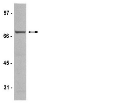
This antibody detects a strong 47 kDa protein, corresponding to the expected molecular mass of GSK3 beta.

Immunogen

Epitope: a.a. 335-349
Rat GSK3 beta synthetic peptide (aa. 335-349), conjugated to KLH. This sequence is 100% identical to human GSK3 beta.

Application

Immunoprecipitation: 12.5 µg/mL dilution of a previous lot was ued in immunoprecipitation on rat brain tissue; immunoprecipitations with tissue extracts other than brain may require higher antibody concentrations.
Optimal working dilutions must be determined by the end user.

Immunohistochemistry(paraffin):
Representative testing from a previous lot.
Optimal Staining of GSK3β Polyclonal Antibody: HeLa Cells
Research Category
Signaling
Research Sub Category
PI3K, Akt, & mTOR Signaling
This AntiI-GSK-3b Antibody, a.a. 335-349 is validated for use in WB, IP, IH(P) for the detection of AntiI-GSK-3b.

Quality

Routinely evaluated by Western Blot on C2C12 lysates.

Western Blot Analysis: 1:500 dilution of this lot detected GSK3 BETA on 10 μg of C2C12 lysates.

Target description

This antibody detects a strong 47 kDa protein.

Linkage

Replaces: AB8687

Physical form

Protein A affinity purified
Purified polyclonal IgG antibody is supplied in PBS, pH 7.2, 50% glycerol and 0.09% sodium azide.

Storage and Stability

Stable at –20 ºC for up to one year from date of receipt. Product may be stored at for 2-4 weeks at 2º-8ºC.
Handling Recommendations: Upon first thaw, and prior to removing the cap, centrifuge the vial and gently mix the solution. Aliquot into microcentrifuge tubes and store at -20°C. Avoid repeated freeze/thaw cycles, which may damage IgG and affect product performance. Note: Variability in freezer temperatures below -20°C may cause glycerol containing solutions to become frozen during storage.

Analysis Note

Control
C2C12 Cell Lysate, Hela cells, rat or mouse brain

Other Notes

Concentration: Please refer to the Certificate of Analysis for the lot-specific concentration.

Disclaimer

Unless otherwise stated in our catalog or other company documentation accompanying the product(s), our products are intended for research use only and are not to be used for any other purpose, which includes but is not limited to, unauthorized commercial uses, in vitro diagnostic uses, ex vivo or in vivo therapeutic uses or any type of consumption or application to humans or animals.

Not finding the right product?  

Try our Product Selector Tool.

Storage Class Code

12 - Non Combustible Liquids

WGK

WGK 2

Flash Point(F)

Not applicable

Flash Point(C)

Not applicable


Certificates of Analysis (COA)

Search for Certificates of Analysis (COA) by entering the products Lot/Batch Number. Lot and Batch Numbers can be found on a product’s label following the words ‘Lot’ or ‘Batch’.

Already Own This Product?

Find documentation for the products that you have recently purchased in the Document Library.

Visit the Document Library

Impact of laminitis on the canonical Wnt signaling pathway in basal epithelial cells of the equine digital laminae.
Wang, L; Pawlak, EA; Johnson, PJ; Belknap, JK; Eades, S; Stack, S; Cousin, H; Black, SJ
Testing null
Zhiyuan Fang et al.
Journal of dairy science, 105(8), 6895-6908 (2022-07-16)
Endoplasmic reticulum (ER) stress can be induced by various stimuli and triggers the unfolded protein response to activate intracellular signaling pathways that are mediated by 3 ER-resident sensors: inositol requiring protein-1α (IRE1α), PKR-like ER kinase (PERK), and activating transcription factor-6
The neuron-specific isoform of glycogen synthase kinase-3beta is required for axon growth.
Casta?o, Zafira, et al.
Journal of Neurochemistry, 113, 117-130 (2010)
Tzyy-Yue Wong et al.
Journal of clinical medicine, 7(11) (2018-11-08)
The dynamics of a living body enables organs to experience mechanical stimulation at cellular level. The human cardiomyocytes cell line provides a source for simulating heart dynamics; however, a limited understanding of the mechanical stimulation effect on them has restricted
Kyung Cheul Shin et al.
Nature communications, 8(1), 1087-1087 (2017-10-24)
Obesity is closely associated with increased adipose tissue macrophages (ATMs), which contribute to systemic insulin resistance and altered lipid metabolism by creating a pro-inflammatory environment. Very low-density lipoprotein receptor (VLDLR) is involved in lipoprotein uptake and storage. However, whether lipid

Our team of scientists has experience in all areas of research including Life Science, Material Science, Chemical Synthesis, Chromatography, Analytical and many others.

Contact Technical Service