Saltar al contenido
Merck
Todas las fotos(2)

Key Documents

MABC592

Sigma-Aldrich

Anti-TRP1/TYRP1 Antibody, clone TA99, Azide Free

clone TA99, 1 mg/mL, from mouse

Sinónimos:

5,6-dihydroxyindole-2-carboxylic acid oxidase, DHICA oxidase, Catalase B, Glycoprotein 75, Melanoma antigen gp75, Tyrosinase-related protein 1, TRP, TRP-1, TRP1

Iniciar sesiónpara Ver la Fijación de precios por contrato y de la organización


About This Item

UNSPSC Code:
12352203
eCl@ss:
32160702
NACRES:
NA.41

biological source

mouse

Quality Level

antibody form

purified immunoglobulin

antibody product type

primary antibodies

clone

TA99, monoclonal

species reactivity

human, mouse

concentration

1 mg/mL

technique(s)

blocking: suitable (interfering antibodies)
flow cytometry: suitable
immunohistochemistry: suitable
immunoprecipitation (IP): suitable
western blot: suitable

isotype

IgG2aκ

NCBI accession no.

UniProt accession no.

shipped in

wet ice

target post-translational modification

unmodified

Gene Information

human ... TYRP1(7306)

General description

TRP1/TYRP1, also known as 5,6-dihydroxyindole-2-carboxylic acid oxidase, DHICA, Catalase B, Glycoprotein 75, Melanoma antigen gp75, or Tyrosinase-related protein 1 (TRP/TRP-1/TRP1), and encoded by the gene TYRP1/CAS2/TYRP/TYRRP, is involved in melanin synthesis and is the enzyme that oxidizes DHICA into indole-5,6-quinone-2-carboxylic acid in the pathway. TRP1/TYRP1 is critical for skin/eye/hair pigmentation. Mutations are associated with Albinism. TRP1/TYRP1 is regulated by the microphthalmia-associated transcription factor (MITF). MITF is a master regulator of pigmentation and a target of β-catenin in Wnt/β-catenin signaling during melanocyte differentiation. TRP1/TYRP1 is also regulated by the microRNA miR-145, and this microRNA is being examined as a possible therapy for hyperpigmentation. TRP1/TYRP1 is expressed as a membrane protein in melanosomes and endosomes in pigment cells.

Immunogen

Whole cells corresponding to human TRP1/TYRP1.

Application

Immunohistochemistry Analysis: A 1:50 dilution from a representative lot detected TRP1/TYRP1 in human melanoma tissue.

Blocking of Interferring Antibodies Analysis: A representative lot from an independent laboratory suppressed the growth of subcutaneous B16 tumors (Patel, D., et al. (2008). Anticancer Res. 28(5A):2679-2686.).

Activity Assay Analysis: A representative lot from an independent laboratory improves anti-tumor efficacy by augmenting systemic CD8+T cell responses to tumor cells (Saenger, Y. M., et al. (2008). Cancer Res. 68(23):9884-9891.).

Immunoprecipiptation Analysis: A representative lot from an independent laboratory immunoprecipiated TRP1/TYRP1 from B16 cell lysate (Srinivasan, R., et al. (2002). Cancer Immun. 19(2):8.).
Research Category
Apoptosis & Cancer
Research Sub Category
Apoptosis - Additional
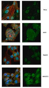
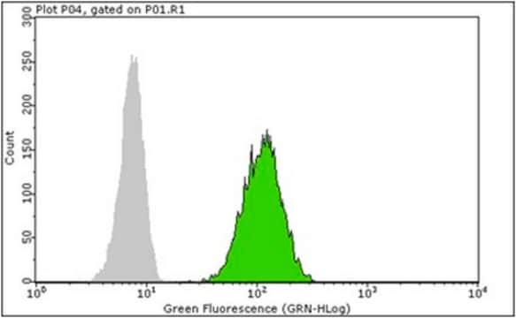
This Anti-TRP1/TYRP1 Antibody, clone TA99, Azide Free is validated for use in western blotting, IHC, blocking of inteferring antibodies, IP & flow cytometry for the detection of TRP1/TYRP1.

Quality

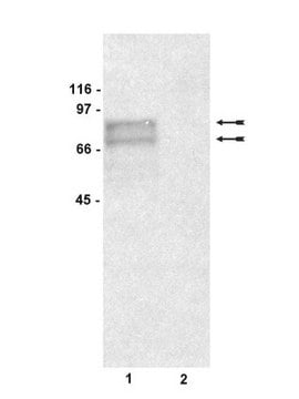
Evaluated by Western Blottng in mouse skin tissue lysate.

Western Blotting Analysis: 1 µg/mL of this antibody detected TRP1/TYRP1 in 10 µg of mouse skin tissue lysate.

Target description

~60 kDa observed

Physical form

Format: Purified
Protein G Purified
Purified mouse monoclonal IgG2aκ in buffer containing PBS without preservatives.

Storage and Stability

Stable for 1 year at -20°C from date of receipt.
Handling Recommendations: Upon receipt and prior to removing the cap, centrifuge the vial and gently mix the solution. Aliquot into microcentrifuge tubes and store at -20°C. Avoid repeated freeze/thaw cycles, which may damage IgG and affect product performance.

Disclaimer

Unless otherwise stated in our catalog or other company documentation accompanying the product(s), our products are intended for research use only and are not to be used for any other purpose, which includes but is not limited to, unauthorized commercial uses, in vitro diagnostic uses, ex vivo or in vivo therapeutic uses or any type of consumption or application to humans or animals.

¿No encuentra el producto adecuado?  

Pruebe nuestro Herramienta de selección de productos.

Storage Class

12 - Non Combustible Liquids

wgk_germany

WGK 2

flash_point_f

Not applicable

flash_point_c

Not applicable


Certificados de análisis (COA)

Busque Certificados de análisis (COA) introduciendo el número de lote del producto. Los números de lote se encuentran en la etiqueta del producto después de las palabras «Lot» o «Batch»

¿Ya tiene este producto?

Encuentre la documentación para los productos que ha comprado recientemente en la Biblioteca de documentos.

Visite la Librería de documentos

Ling Jiang et al.
Journal of cellular physiology, 234(12), 22799-22808 (2019-05-23)
Our previous study found that Ganoderma lucidum polysaccharide (GLP), bioactive ingredients from Ganoderma lucidum, protected fibroblasts from photoaging. However, whether GLP can affect melanogenesis in melanocytes through regulating paracrine mediators that secreted by keratinocytes and fibroblasts is unclear. We aimed
C L Slingluff et al.
Cancer immunology, immunotherapy : CII, 48(12), 661-672 (2001-02-07)
Melanoma-reactive HLA-A x 0201-restricted cytotoxic T lymphocyte (CTL) lines generated in vitro lyse autologous and HLA-matched allogeneic melanoma cells and recognize multiple shared peptide antigens from tyrosinase, MART-1, and Pme117/gp100. However, a subset of melanomas fail to be lysed by
Shuanghai Hu et al.
Journal of cellular physiology, 234(5), 7330-7340 (2018-10-27)
Ultraviolet (UV)-induced pigmentation is very common in clinical practice, but the current treatments are rarely effective, accompanied by some side effects. Ganoderma lucidum polysaccharide (GLP) is a natural antioxidant with no toxic side effects, which can antagonize UVB-induced fibroblast photo
Shiyao Pei et al.
The Journal of investigative dermatology, 140(1), 152-163 (2019-07-06)
The long noncoding RNA UCA1 was first discovered in bladder cancer and is known to regulate the proliferation and migration of melanoma. However, its role in melanogenesis is unclear. In this study, we aimed to explore the role and mechanism
Li-Ping Liu et al.
Cell reports, 27(2), 455-466 (2019-04-11)
Induced pluripotent stem cells (iPSCs) are a promising melanocyte source as they propagate indefinitely and can be established from patients. However, the in vivo functions of human iPSC-derived melanocytes (hiMels) remain unknown. Here, we generated hiMels from vitiligo patients using a three-dimensional

Nuestro equipo de científicos tiene experiencia en todas las áreas de investigación: Ciencias de la vida, Ciencia de los materiales, Síntesis química, Cromatografía, Analítica y muchas otras.

Póngase en contacto con el Servicio técnico