
09-403
Anti-TKS5 (SH3 #1) Antibody
from rabbit
Sinónimos:
Adaptor protein TKS5, Five SH3 domain-containing protein, SH3 and PX domains 2A, SH3 multiple domains 1, SH3 multiple domains protein 1, five SH3 domains
About This Item
Productos recomendados


origen biológico
rabbit
Nivel de calidad
forma del anticuerpo
purified antibody
tipo de anticuerpo
primary antibodies
clon
polyclonal
reactividad de especies
mouse, human
técnicas
immunocytochemistry: suitable
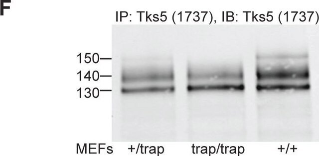
immunoprecipitation (IP): suitable
western blot: suitable
Nº de acceso NCBI
Nº de acceso UniProt
Condiciones de envío
wet ice
modificación del objetivo postraduccional
unmodified
Información sobre el gen
human ... SH3PXD2A(9644)
mouse ... Sh3Pxd2A(14218)
Descripción general
Especificidad
Inmunógeno
Aplicación
Cell Structure
Cytoskeletal Signaling
Calidad
Descripción de destino
Forma física
Almacenamiento y estabilidad
Nota de análisis
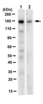
Western Blot::
NIH-3T3 (100% confluent) lysate
Immunocytochemistry:
Mouse 3T3-Src(Y527F) cells
Otras notas
Cláusula de descargo de responsabilidad
¿No encuentra el producto adecuado?
Pruebe nuestro Herramienta de selección de productos.
Código de clase de almacenamiento
12 - Non Combustible Liquids
Clase de riesgo para el agua (WGK)
WGK 1
Punto de inflamabilidad (°F)
Not applicable
Punto de inflamabilidad (°C)
Not applicable
Certificados de análisis (COA)
Busque Certificados de análisis (COA) introduciendo el número de lote del producto. Los números de lote se encuentran en la etiqueta del producto después de las palabras «Lot» o «Batch»
¿Ya tiene este producto?
Encuentre la documentación para los productos que ha comprado recientemente en la Biblioteca de documentos.
Nuestro equipo de científicos tiene experiencia en todas las áreas de investigación: Ciencias de la vida, Ciencia de los materiales, Síntesis química, Cromatografía, Analítica y muchas otras.
Póngase en contacto con el Servicio técnico