Skip to Content
Merck
All Photos(1)

Key Documents

203A-1

Sigma-Aldrich

Alpha-Fetoprotein Rabbit Polyclonal Antibody

Sign Into View Organizational & Contract Pricing


About This Item

UNSPSC Code:
12352203
NACRES:
NA.41

biological source

rabbit

Quality Level

100
500

conjugate

unconjugated

antibody form

Ig fraction of antiserum

antibody product type

primary antibodies

clone

polyclonal

description

For In Vitro Diagnostic Use in Select Regions (See Chart)

form

buffered aqueous solution

species reactivity

human

packaging

vial of 0.1 mL concentrate (203A-14)
vial of 0.5 mL concentrate (203A-15)
bottle of 1.0 mL predilute (203A-17)
vial of 1.0 mL concentrate (203A-16)
bottle of 7.0 mL predilute (203A-18)

manufacturer/tradename

Cell Marque

technique(s)

immunohistochemistry (formalin-fixed, paraffin-embedded sections): 1:50-1:200

control

fetal liver

shipped in

wet ice

storage temp.

2-8°C

visualization

cytoplasmic

General description

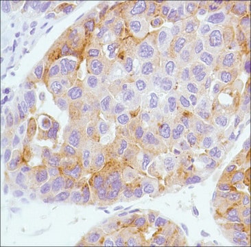
Alpha-fetoprotein (AFP) is a fetal tumor-associated polypeptide of the albuminoid gene family that binds and transports molecules in addition to many other proposed functions. This secretory protein is produced by the fetal yolk sac and liver but is found only in trace amounts in adult liver. AFP expression by immunohistochemistry is seen in hepatocellular carcinoma (HCC).
Positive staining with anti-AFP is seen in hepatocytes of the fetal liver and hepatoma. Since only traces of AFP are found in adult serum, elevated levels suggest either a benign or malignant lesion of the liver, a yolk sac carcinoma, or one of a few other tumors. Correspondingly, in conjunction with elevated serum levels, AFP has been immunohistochemically demonstrated in yolk sac tumors of gonadal and extragonadal sites, in hepatic malignancies, and in a few other neoplasms. The antigen is not denatured by most fixatives.

Quality


IVD

IVD

IVD

RUO

Linkage

Alpha-Fetoprotein Positive Control Slides, Product No. 203S, are available for immunohistochemistry (formalin-fixed, paraffin-embedded sections).

Physical form

Solution in Tris Buffer, pH 7.3-7.7, with 1% BSA and <0.1% Sodium Azide

Preparation Note

Download the IFU specific to your product lot and formatNote: This requires a keycode which can be found on your packaging or product label.

Other Notes

For Technical Service please contact: 800-665-7284 or email: service@cellmarque.com

Legal Information

Cell Marque is a trademark of Merck KGaA, Darmstadt, Germany

Not finding the right product?  

Try our Product Selector Tool.


Certificates of Analysis (COA)

Search for Certificates of Analysis (COA) by entering the products Lot/Batch Number. Lot and Batch Numbers can be found on a product’s label following the words ‘Lot’ or ‘Batch’.

Already Own This Product?

Find documentation for the products that you have recently purchased in the Document Library.

Visit the Document Library

Manual of Diagnostic Antibodies for Immunohistology, 9-10 (1999)
S Peyrol et al.
Digestion, 18(5-6), 351-370 (1978-01-01)
5 human cases of hepatoma have been chosen with respect to their different seric alpha-fetoprotein (alpha-FP) level and histological characters. Cells producing alpha-FP have been studied with specific horseradish-peroxidase-labelled immunoglobulins. Ultrastructural examination shows that alpha-FP is present in the cytoplasm
S H Tsung
Archives of pathology & laboratory medicine, 101(11), 572-574 (1977-11-01)
Immunofluorescence staining was applied to tissue with primary nonhepatic malignancies and/or their metastatic nodules in the liver. Tumor cells from the primary sites of two patients with carcinoma of the pancreas and one patient with Hodgkin's disease were found to
L M Roth et al.
Cancer, 37(2), 812-820 (1976-02-01)
Fourteen cases of yolk sac carcinoma, 10 occurring in gonadal, and four in extragonadal sites, seen at the Indiana University Hospitals from 1949 to 1974, were analyzed with respect to pathologic features, laboratory findings, and clinical course. Their histologic appearance
Z D Goodman et al.
Cancer, 55(1), 124-135 (1985-01-01)
Combined hepatocellular-cholangiocarcinoma is a rare form of primary liver cancer showing features of both hepatocellular and biliary epithelial differentiation. In a review of 24 cases of this tumor, three histologic types were encountered. Four cases were Type I or "collision

Our team of scientists has experience in all areas of research including Life Science, Material Science, Chemical Synthesis, Chromatography, Analytical and many others.

Contact Technical Service