Skip to Content
Merck
All Photos(8)

Documents

A2547

Sigma-Aldrich

Monoclonal Anti-Actin, α-Smooth Muscle

clone 1A4, ascites fluid

Synonym(s):

Alpha Smooth Muscle Actin Antibody - Monoclonal Anti-Actin, α-Smooth Muscle, Alpha Smooth Muscle Actin Antibody Sigma, Anti-Alpha Smooth Muscle Actin Antibody, SMA

Sign Into View Organizational & Contract Pricing


About This Item

UNSPSC Code:
12352203
NACRES:
NA.41

biological source

mouse

conjugate

unconjugated

antibody form

ascites fluid

antibody product type

primary antibodies

clone

1A4, monoclonal

mol wt

antigen ~42 kDa

contains

15 mM sodium azide

species reactivity

human, mouse, rat, chicken, frog, canine, rabbit, guinea pig, goat, bovine, sheep, snake

technique(s)

immunohistochemistry (formalin-fixed, paraffin-embedded sections): suitable using smooth muscle cells
immunohistochemistry (frozen sections): suitable using smooth muscle cells
indirect immunofluorescence: 1:400 using blood vessels in sections of human appendix
western blot: suitable using smooth muscle cells

isotype

IgG2a

UniProt accession no.

application(s)

research pathology

shipped in

dry ice

storage temp.

−20°C

target post-translational modification

unmodified

Gene Information

human ... ACTA2(59)
mouse ... Acta2(11475)
rat ... Acta2(81633)

Looking for similar products? Visit Product Comparison Guide

Related Categories

General description

Actins are cytoskeletal proteins that regulate cell motility. Several scaffolding, signaling and actin-binding proteins regulate the interconversions between filamentous F-actins and monomeric G-actins. Cellular actins of various species have very similar immunological and physical properties. Hence, the use of monoclonal antibody to actin provides a specific and useful tool for studying the localization, structure and function of actin proteins. Actin 2 (ACTA2), the smooth muscle actin is encoded on human chromosome 10q23.31.
Mouse monoclonal anti-actin, α-smooth muscle antibody binds to actin from human, mouse, rat, bovine, chicken, frog, goat, guinea pig, rabbit, dog, sheep, and snake.
Monoclonal Anti-α Smooth Muscle Actin (mouse IgG2a isotype) is derived from the hybridoma produced by the fusion of mouse myeloma cells and splenocytes from an immunized mouse. The NH2 terminal synthetic decapeptide of α-smooth muscle actin coupled to keyhole limpet hemocyanin (KLH) was used as the immunogen. The isotype is determined using Sigma ImmunoTypeTM Kit (Sigma Stock No. ISO-1) and by a double diffusion immunoassay using Mouse Monoclonal Antibody Isotyping Reagents (Sigma Stock No. ISO-2).
The antibody (also known as anti-α-Sm-1) is specific for the single isoform of α-smooth muscle actin. It reacts specifically with α-smooth muscle actin in immunoblotting assays and labels smooth muscle cells in frozen or formalin-fixed, paraffin-embedded tissue sections.

Specificity

Monoclonal Anti-α Smooth Muscle Actin is specific for the single isoform α-actin. It reacts specifically with a smooth muscle actin using immunoblotting and indirect immunofluorescent labeling of formalin-fixed, paraffin embedded or frozen tissue sections. The antibody can be used for immunocytochemical localization of the α smooth muscle actin by means of immunoperoxidase, immunofluorescent, ELISA or immunoblot techniques. The antibody reacts with human, mouse, rat, bovine, chicken, frog, goat, guinea pig, rabbit, dog, sheep, and snake.

Immunogen

N-terminal synthetic decapeptide of α-smooth muscle actin.

Application

Immunocytochemistry was performed on smooth muscle cells from bovine aortas using the monoclonal anti-ACTA2 antibody. Cells were first grown on glass cover slips and fixed in 50% acetone/EtOH for 10 minutes at 4 degrees.
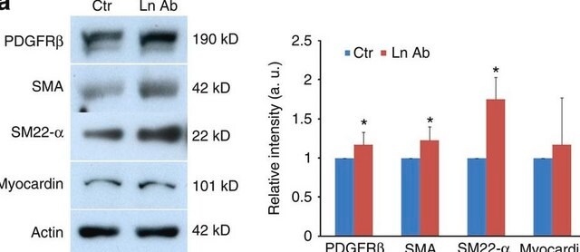
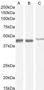
Mouse monoclonal anti-actin, α-smooth muscle antibody has been used for western blot assays using whole kidney homogenates in human anaplastic thyroid cancer cells. The product has also been used for immunohistochemical assays using mice tissue sections in liver sections and uterine sections.
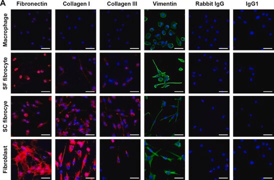
Monoclonal Anti-Actin, α-Smooth Muscle antibody has been used in the detection of actin 2 in breast fibroblasts using immunofluorescence staining.
Paraffin embedded sections of rat testis tissue grafts were immunohistochemically stained with mouse monoclonal anti-smooth muscle actin.
IHC analysis of x-gal stained muouse cardiac tissue was performed using the primary antibody, mouse monoclonal anti-smooth muscle actin to identify myofibroblasts.

Biochem/physiol Actions

Actin is one of the most conserved eukaryotic proteins, it is expressed in mammals and birds as six isoforms characterized by electrophoresis and amino acid sequence analysis. Four of the six represent differentiation markers of muscle tissues. Actin isoforms show >90% overall sequence homology, but only 50-60% homology in their 18 NH2-terminal residues. The NH2-terminal region of actin appears to be a major antigenic region, and may be involved in the interaction of actin with other proteins such as myosin. It has been shown that the relative proportion of actin isoforms are different in smooth muscles of different organs and change within the same population of smooth muscle cells during development, pathological situations and different culture conditions.
Mutations in actin 2 is associated with dysfunction of smooth muscles in arteries and is implicated in coronary heart disease. Mutations in ACTA2 contributes to tear and enlargement of aorta in thoracic aortic aneurysms (TAAD).

Physical form

The antibody is provided as ascites fluid containing 15mM sodium azide as a preservative.

Storage and Stability

For continuous use, store at 2-8 °C for up to one month. For extended storage, solution may be frozen in working aliquots. Repeated freezing and thawing is not recommended. If slight turbidity occurs upon prolonged storage, clarify by centrifugation before use.

Other Notes

To view an Actin antibody selection guide, please visit www.sigmaaldrich.com/actin.

Disclaimer

Unless otherwise stated in our catalog or other company documentation accompanying the product(s), our products are intended for research use only and are not to be used for any other purpose, which includes but is not limited to, unauthorized commercial uses, in vitro diagnostic uses, ex vivo or in vivo therapeutic uses or any type of consumption or application to humans or animals.

Not finding the right product?  

Try our Product Selector Tool.

Storage Class Code

10 - Combustible liquids

WGK

WGK 1

Flash Point(F)

Not applicable

Flash Point(C)

Not applicable


Certificates of Analysis (COA)

Search for Certificates of Analysis (COA) by entering the products Lot/Batch Number. Lot and Batch Numbers can be found on a product’s label following the words ‘Lot’ or ‘Batch’.

Already Own This Product?

Find documentation for the products that you have recently purchased in the Document Library.

Visit the Document Library

Natalie C Direkze et al.
Cancer research, 64(23), 8492-8495 (2004-12-03)
The role of myofibroblasts in tissue repair and fibrosis is well documented, but the source of these myofibroblasts is unclear. There is evidence of a circulating population of fibrocytes that can home to areas of injury and contribute to myofibroblast
Yijie Jia et al.
Molecular therapy : the journal of the American Society of Gene Therapy, 27(8), 1452-1466 (2019-06-19)
Albumin absorbed by renal tubular epithelial cells induces inflammation and plays a key role in promoting diabetic kidney disease (DKD) progression. Macrophages are prominent inflammatory cells in the kidney, and their role there is dependent on their phenotypes. However, whether
Differential changes in 15-hydroxyprostaglandin dehydrogenase and prostaglandin H synthase (types I and II) in human pregnant myometrium.
Giannoulias D, et al.
The Journal of Clinical Endocrinology and Metabolism, 87(3), 1345-1352 (2002)
Indirect and direct revascularization of ACTA2 cerebral arteriopathy: feasibility of the superficial temporal artery to anterior cerebral artery bypass with posterior auricular artery interposition graft: case report.
Rutledge WC, et al.
Journal of Neurosurgery. Pediatrics, 18(3), 339-343 (2016)
Thrombospondin-1 Silencing Down-Regulates Integrin Expression Levels in Human Anaplastic Thyroid Cancer Cells with BRAFV600E: New Insights in the Host Tissue Adaptation and Homeostasis of Tumor Microenvironment.
Duquette M, et al.
Frontiers in Endocrinology, 4, 189-189 null

Our team of scientists has experience in all areas of research including Life Science, Material Science, Chemical Synthesis, Chromatography, Analytical and many others.

Contact Technical Service