Skip to Content
Merck
All Photos(1)

Documents

CBL412X

Sigma-Aldrich

Anti-Tubulin β-III Antibody, clone TU-20, Alexa Fluor488 Conjugated

clone TU-20, from mouse, ALEXA FLUOR 488

Synonym(s):

Tubulin beta-4, Tubulin beta-III, tubulin, beta 3, tubulin, beta 4

Sign Into View Organizational & Contract Pricing


About This Item

UNSPSC Code:
12352203
eCl@ss:
32160702
NACRES:
NA.41

biological source

mouse

Quality Level

conjugate

ALEXA FLUOR 488

antibody form

purified immunoglobulin

antibody product type

primary antibodies

clone

TU-20, monoclonal

species reactivity

human, mouse, pig, bovine, primate, rat

technique(s)

immunocytochemistry: suitable
immunofluorescence: suitable
immunohistochemistry: suitable
immunoprecipitation (IP): suitable
western blot: suitable

isotype

IgG1

NCBI accession no.

UniProt accession no.

shipped in

wet ice

target post-translational modification

unmodified

Gene Information

human ... TUBB3(10381)

General description

Tubulin is one of several members of a small family of globular proteins which polymerize to form cytoskeletal structures called microtubules. The most common members of the tubulin family are α-tubulin and β-tubulin. Tubulin dimers, typically composed of α- and β-tubulin subunits, assemble onto microtubules forming large polymers. β-III tubulin is a unique microtubule subunit that is found expressed almost exclusively in neurons. Due to the unique expression pattern of β-III tubulin it is often utilized as a marker to identify neuronal cells from other brain cells such as glia which do not express it. β-III tubulin also serves as a cytoarchitectural marker since it is primarily found within neurite extensions.

Specificity

Reactivity with other species has not been determined.
This antibody reacts with the C-terminus of the beta-III isoform of tubulin, which is known to be neuron specific. Does not identify beta tubulin found in glial cells.

Immunogen

Epitope: C-terminus

Application

Detect Tubulin β-III using this Anti-Tubulin β-III Antibody, clone TU-20, Alexa Fluor488 Conjugated validated for use in IC, IF, WB, IH & IP.
Immunohistochemistry: 5-10 µg/mL on frozen brain tissue.
Although this conjugated antibody will work for Western blotting and Immunoprecipitation, we recommend catalog number CBL412 if these are your primary applications.
Optimal working concentration must be determined by end user.
Research Category
Neuroscience
Research Sub Category
Neurofilament & Neuron Metabolism

Quality

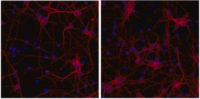
Routinely tested cortical neurons in Immunocytochemistry.
Immunocytochemical Analysis: 1:250 dilution of this lot detected β-III tubulin in rat cortex cell culture.

Target description

50 kDa

Physical form

Protein G Purified
Purified immunoglobulin in 0.02M Phosphate buffer, 0.25M NaCl containing 0.05% sodium azide.

Storage and Stability

Stable for 6 months at 2-8°C from date of receipt.

Analysis Note

Control
Rat Cortex Cells.

Other Notes

Concentration: Please refer to the Certificate of Analysis for the lot-specific concentration.

Legal Information

ALEXA FLUOR is a trademark of Life Technologies

Disclaimer

Unless otherwise stated in our catalog or other company documentation accompanying the product(s), our products are intended for research use only and are not to be used for any other purpose, which includes but is not limited to, unauthorized commercial uses, in vitro diagnostic uses, ex vivo or in vivo therapeutic uses or any type of consumption or application to humans or animals.

Not finding the right product?  

Try our Product Selector Tool.

Storage Class Code

12 - Non Combustible Liquids

WGK

WGK 2

Flash Point(F)

Not applicable

Flash Point(C)

Not applicable


Certificates of Analysis (COA)

Search for Certificates of Analysis (COA) by entering the products Lot/Batch Number. Lot and Batch Numbers can be found on a product’s label following the words ‘Lot’ or ‘Batch’.

Already Own This Product?

Find documentation for the products that you have recently purchased in the Document Library.

Visit the Document Library

Shin-Ichiro Sekine et al.
Stem cell research, 24, 40-43 (2017-10-17)
Idiopathic basal ganglia calcification (IBGC), also known as Fahr disease or primary familial brain calcifications (PFBC), is a rare neurodegenerative disorder characterized by calcium deposits in basal ganglia and other brain regions, causing neuropsychiatric and motor symptoms. We established human
Elena Romano et al.
Stem cell research, 57, 102580-102580 (2021-10-24)
Human induced pluripotent stem cells (iPSCs) have great promise in regenerative medicine. However, several limitations, including immune-incompatibility, have raised concerns regarding their clinical application. Recent studies have shown that human iPSCs and their derivatives lose their immunogenicity when major histocompatibility
Angela R Dixon et al.
Toxicology in vitro : an international journal published in association with BIBRA, 29(3), 564-574 (2015-01-03)
With m-Dinitrobenzene (m-DNB) as a selected model neurotoxicant, we demonstrate how to assess neurotoxicity, using morphology based measurement of neurite degeneration, in a conventional "full-contact" and a modern "restricted-contact" co-culture of rat cortical neurons and astrocytes. In the "full-contact" co-culture
Courtney M Tate et al.
PloS one, 10(4), e0125697-e0125697 (2015-04-29)
Bone morphogenetic proteins (BMPs), members of the TGF-β superfamily, have numerous biological activities including control of growth, differentiation, and vascular development. Using an in vitro co-culture endothelial cord formation assay, we investigated the role of a BMP7 variant (BMP7v) in

Our team of scientists has experience in all areas of research including Life Science, Material Science, Chemical Synthesis, Chromatography, Analytical and many others.

Contact Technical Service