Accéder au contenu
Merck
Toutes les photos(5)

Key Documents

A4200

Sigma-Aldrich

Monoclonal Anti-γ-Adaptin antibody produced in mouse

clone 100/3, ascites fluid

Synonyme(s) :

Anti-AP-1

Se connecterpour consulter vos tarifs contractuels et ceux de votre entreprise/organisme


About This Item

Numéro MDL:
Code UNSPSC :
12352203
Nomenclature NACRES :
NA.41

Source biologique

mouse

Conjugué

unconjugated

Forme d'anticorps

ascites fluid

Type de produit anticorps

primary antibodies

Clone

100/3, monoclonal

Poids mol.

antigen 104 kDa

Contient

15 mM sodium azide

Espèces réactives

bovine, human, monkey

Ne doit pas réagir avec

mouse, rat

Technique(s)

electron microscopy: suitable
immunoprecipitation (IP): suitable
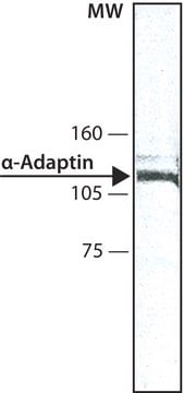
western blot: 1:100 using bovine brain extract

Isotype

IgG2b

Numéro d'accès UniProt

Conditions d'expédition

dry ice

Température de stockage

−20°C

Modification post-traductionnelle de la cible

unmodified

Informations sur le gène

human ... AP1G1(164)
mouse ... Ap1g1(11765)
rat ... Ap1g1(171494)

Description générale

Clathrin-coated vesicle populations contain the adaptor complexes AP-1 and AP-2, also known as HA-I (HA1) adaptor and HA-II (HA2) adaptor or Assemble Protein 1 (AP1) and Assemble Protein 2 (AP2), respectively. Clathrin-coated membranes at or originating from the plasma membrane contain the AP-2 adaptor, while the AP-1 adaptor appears to be largely restricted to clathrin-coated membranes of the trans-Golgi network. Both the Golgi-associated AP-1 adaptor and the plasma-membrane associated AP-2 adaptor are heterotetrameric protein complexes. AP-1 adaptor consists of four subunits termed b1 (formerly b′, 110 kDa), g (100 kDa), m1 (47 kDa), and s1 (20 kDa). b1 and g subunits (b1 and g adaptins) from neuronal sources behave in standard SDS-PAGE like 115 kDa and 104 kDa polypeptides, respectively.

Spécificité

Mouse monoclonal clone 100/3 anti-g-Adaptin antibody reacts in immunoblotting with the 104 kDa polypeptide of the Golgi adaptor complex AP-1. It reacts with polypeptides of ~100 kDa in bovine liver, human heart fibroblasts, and Madin-Darby bovine kidney cultured cells (MDBK), but not with any components in the 110-115 kDa range from these sources or from neuroblastoma or astrocytes. This suggests that the 110 and 115 kDa polypeptides may be specific variants occurring only in some cell types of brain. The antibody does not recognize the γ-subunit in rodents (rat and mouse).

Immunogène

AP-1 adaptor from bovine brain.

Application

Applications in which this antibody has been used successfully, and the associated peer-reviewed papers, are given below.
Immuno-electron microscopy (1 paper)
Immunofluorescence (1 paper)
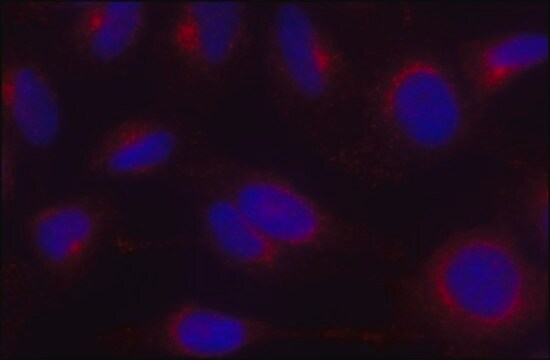
Immunofluorescence of Hela cells was performed using monoclonal anti-AP1 (clone 100/3) as the primary antibody.
In immuno­fluorescence using MDBK cells, human heart fibroblasts, and African green monkey kidney cells (Vero), the binding of antibody appears to be largely confined to the trans-Golgi network. The antibody has been used for studies on the effects of Brefeldin A, a substance causing rapid redistribution of coat proteins associated with the clathrin-coated vesicles that bud from the trans-Golgi network. The antibody is also useful for the immunoaffinity purification of the Golgi adaptor complex AP-1.
Mouse monoclonal anti-gamma-adaptin antibody can be used for western blot (1:100, using bovine brain extract), immunoprecipitation and electron microscopy applications.

Forme physique

The product is provided as ascites fluid with 0.1% sodium azide as a preservative.

Clause de non-responsabilité

Unless otherwise stated in our catalog or other company documentation accompanying the product(s), our products are intended for research use only and are not to be used for any other purpose, which includes but is not limited to, unauthorized commercial uses, in vitro diagnostic uses, ex vivo or in vivo therapeutic uses or any type of consumption or application to humans or animals.

Vous ne trouvez pas le bon produit ?  

Essayez notre Outil de sélection de produits.

Code de la classe de stockage

13 - Non Combustible Solids

Classe de danger pour l'eau (WGK)

WGK 1

Point d'éclair (°F)

Not applicable

Point d'éclair (°C)

Not applicable


Certificats d'analyse (COA)

Recherchez un Certificats d'analyse (COA) en saisissant le numéro de lot du produit. Les numéros de lot figurent sur l'étiquette du produit après les mots "Lot" ou "Batch".

Déjà en possession de ce produit ?

Retrouvez la documentation relative aux produits que vous avez récemment achetés dans la Bibliothèque de documents.

Consulter la Bibliothèque de documents

Wiebke Stahlschmidt et al.
The Journal of biological chemistry, 289(8), 4906-4918 (2014-01-11)
Clathrin plays important roles in intracellular membrane traffic including endocytosis of plasma membrane proteins and receptors and protein sorting between the trans-Golgi network (TGN) and endosomes. Whether clathrin serves additional roles in receptor recycling, degradative sorting, or constitutive secretion has
Brogan Yarzabek et al.
eLife, 7 (2018-07-11)
The highly polymorphic human leukocyte antigen (HLA) class I molecules present peptide antigens to CD8+ T cells, inducing immunity against infections and cancers. Quality control mediated by peptide loading complex (PLC) components is expected to ensure the cell surface expression
Karin Håberg et al.
Journal of cell science, 121(Pt 9), 1495-1505 (2008-04-16)
SNX9, SNX18 and SNX30 constitute a separate subfamily of PX-BAR-containing sorting nexin (SNX) proteins. We show here that most tissues express all three paralogs, and immunoprecipitation and immunofluorescence experiments demonstrated that the SNX9-family proteins act as individual entities in cells.
Jorge Cancino et al.
Molecular biology of the cell, 18(12), 4872-4884 (2007-09-21)
The epithelial-specific adaptor AP1B sorts basolateral plasma membrane (PM) proteins in both biosynthetic and recycling routes, but the site where it carries out this function remains incompletely defined. Here, we have investigated this topic in Fischer rat thyroid (FRT) epithelial
Nicole A Ducharme et al.
Cellular logistics, 1(2), 57-68 (2011-06-21)
The Rab11 Family Interacting Proteins (Rab11-FIPs) are hypothesized to regulate sequential steps in the apical recycling and transcytotic pathways of polarized epithelial cells. Previous studies have suggested that Rab11-FIP proteins assemble into multi-protein complexes regulating plasma membrane recycling. Rab11-FIP2 interacts

Notre équipe de scientifiques dispose d'une expérience dans tous les secteurs de la recherche, notamment en sciences de la vie, science des matériaux, synthèse chimique, chromatographie, analyse et dans de nombreux autres domaines..

Contacter notre Service technique