Skip to Content
Merck
All Photos(1)

Key Documents

406M-1

Sigma-Aldrich

MUC4 (8G7) Mouse Monoclonal Antibody

Sign Into View Organizational & Contract Pricing


About This Item

UNSPSC Code:
12352203
NACRES:
NA.41

biological source

mouse

Quality Level

100
500

conjugate

unconjugated

antibody form

culture supernatant

antibody product type

primary antibodies

clone

8G7, monoclonal

description

For In Vitro Diagnostic Use in Select Regions (See Chart)

form

buffered aqueous solution

species reactivity

human

packaging

vial of 0.1 mL concentrate (406M-14)
vial of 0.5 mL concentrate (406M-15)
bottle of 1.0 mL predilute (406M-17)
vial of 1.0 mL concentrate (406M-16)
bottle of 7.0 mL predilute (406M-18)

manufacturer/tradename

Cell Marque

technique(s)

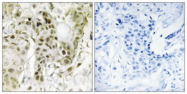
immunohistochemistry (formalin-fixed, paraffin-embedded sections): 1:10-1:50

isotype

IgG1κ

control

colon, colorectal adenocarcinoma, pancreatic ductal adenocarcinoma

shipped in

wet ice

storage temp.

2-8°C

visualization

cytoplasmic

General description

Mucin 4 (MUC4) is a transmembranous glycoprotein. MUC4 is expressed in the cytoplasm of certain epithelial surfaces, such as colonic, breast, and lung epithelia. An abnormal expression of MUC4 has been reported in various carcinomas of the colon, pancreas, breast, and ovaries.MUC4 is highly expressed in the vast majority of human pancreatic neoplasms, such as intraductal papillary mucinous neoplasia of the pancreas, pancreatic intraepithelial neoplasms and pancreatic ductal adenocarcinoma; however, it is not detected in the normal pancreas or in chronic pancreatitis.MUC4 is not expressed in normal bile ducts, but it can be overexpressed in intrahepatic cholangiocarcinoma and bile duct cholangiocarcinoma. MUC4 overexpression has been reported in low-grade fibromyxoid sarcoma (LGFMS). In this study a large cohort of LGFMS, all with FUS gene rearrangement confirmed by FISH, showed cytoplasmic staining for MUC4, usually in a strong and diffuse manner. Similarly, strong, diffuse cytoplasmic staining for MUC4 was identified in 78% (32/41) of cases of sclerosing epithelioid fibrosarcoma. MUC4 expression is also detected in the glandular component of biphasic synovial sarcomas (90%). Focal staining, usually interpreted as only scattered cells, was also seen in a subset of ossifying fibromyxoid tumors (29%), epithelioid GISTs (20%) and myoepithelial carcinomas (10%); whereas all other epithelioid soft tissue tumors—including clear cell sarcoma, epithelioid sarcoma, epithelioid hemangiosarcoma, PEComa and melanoma—were negative. It should be noted that various carcinomas may express MUC4, and therefore additional keratins or lineage-specific markers may be needed to exclude this possibility in some cases.

Quality


IVD

IVD

IVD

RUO

Linkage

MUC4 Positive Control Slides, Product No. 406S, are available for immunohistochemistry (formalin-fixed, paraffin-embedded sections).

Physical form

Solution in Tris Buffer, pH 7.3-7.7, with 1% BSA and <0.1% Sodium Azide

Preparation Note

Download the IFU specific to your product lot and formatNote: This requires a keycode which can be found on your packaging or product label.

Other Notes

For Technical Service please contact: 800-665-7284 or email: service@cellmarque.com

Legal Information

Cell Marque is a trademark of Merck KGaA, Darmstadt, Germany

Not finding the right product?  

Try our Product Selector Tool.


Certificates of Analysis (COA)

Search for Certificates of Analysis (COA) by entering the products Lot/Batch Number. Lot and Batch Numbers can be found on a product’s label following the words ‘Lot’ or ‘Batch’.

Already Own This Product?

Find documentation for the products that you have recently purchased in the Document Library.

Visit the Document Library

Leona A Doyle et al.
The American journal of surgical pathology, 36(10), 1444-1451 (2012-09-18)
Sclerosing epithelioid fibrosarcoma (SEF) is a rare aggressive fibroblastic neoplasm composed of cords of epithelioid cells embedded in a dense collagenous stroma. The reported immunophenotype of SEF is nonspecific. Some SEF cases show morphologic and molecular overlap with low-grade fibromyxoid
N Moniaux et al.
British journal of cancer, 91(9), 1633-1638 (2004-10-21)
Mucins are members of an expanding family of large multifunctional glycoproteins. Pancreatic mucins have important biological functions, including the protection, lubrication, and moisturisation of the surfaces of epithelial tissues lining ductal structures within the pancreas. Several lines of evidence support
Suguru Yonezawa et al.
Journal of hepato-biliary-pancreatic sciences, 17(2), 108-124 (2009-09-30)
Mucins are high molecular weight glycoproteins that play important roles in carcinogenesis and tumor invasion. We have described, for the first time, that pancreatic ductal adenocarcinomas (PDACs) with an aggressive behavior and a poor outcome expressed MUC1 (pan-epithelial membrane-associated mucin)
Leona A Doyle et al.
The American journal of surgical pathology, 35(5), 733-741 (2011-03-19)
Low-grade fibromyxoid sarcoma (LGFMS) is a distinctive fibroblastic neoplasm that is characterized by alternating collagenous and myxoid areas, deceptively bland spindle cell morphology, a whorling architecture, and a t(7;16) translocation involving FUS and CREB3L2. Owing to variable morphology and a
Nicolas Moniaux et al.
The journal of histochemistry and cytochemistry : official journal of the Histochemistry Society, 52(2), 253-261 (2004-01-20)
We have previously cloned the full-length cDNA (approximately 28 Kb) and established the complete genomic organization (25 exons/introns over 100 kb) of the human MUC4 mucin. This large molecule is predicted to protrude over 2 microm above the cell surface

Our team of scientists has experience in all areas of research including Life Science, Material Science, Chemical Synthesis, Chromatography, Analytical and many others.

Contact Technical Service