Accéder au contenu
Merck
Toutes les photos(3)

Key Documents

ABC99

Sigma-Aldrich

Anti-SFTPC Antibody

from rabbit, purified by affinity chromatography

Synonyme(s) :

Pulmonary surfactant-associated protein C, SP-C, Pulmonary surfactant-associated proteolipid SPL(Val), SP5

Se connecterpour consulter vos tarifs contractuels et ceux de votre entreprise/organisme


About This Item

Code UNSPSC :
12352203
eCl@ss :
32160702
Nomenclature NACRES :
NA.41

Source biologique

rabbit

Niveau de qualité

Forme d'anticorps

affinity isolated antibody

Type de produit anticorps

primary antibodies

Clone

polyclonal

Produit purifié par

affinity chromatography

Espèces réactives

mouse, rat, human

Technique(s)

immunohistochemistry: suitable (paraffin)
western blot: suitable

Numéro d'accès NCBI

Numéro d'accès UniProt

Conditions d'expédition

wet ice

Modification post-traductionnelle de la cible

unmodified

Informations sur le gène

human ... SFTPC(6440)

Description générale

SFTPC, also known as SFTP2 and Pulmonary surfactant-associated protein C, is a complex mixture of phospholipids and proteins secreted from airway epithelial cells (type II cells) in alveoli which reduces the surface tension at the alveolar air liquid interface, providing alveolar stability necessary for normal ventilation. Four distinct proteins isolated from pulmonary surfactant are termed surfactant proteins A, B, C, and D. SP A (28 - 36 kDa) and SP D (43 kDa) are collagenous carbohydrate binding proteins, whereas SP B (8 - 9 kDa) and SP C (4 kDa) are non collagenous hydrophobic proteins. SP C is a fragment of a much larger precursor protein of 21 kDa. The precursor contains an exceedingly hydrophobic region of 34 amino acids that comprises most of the mature SP C, and a unique poly-valine domain.

Immunogène

Epitope: N-terminus
KLH-conjugated linear peptide corresponding to the N-terminus of human SFTPC.

Application

Detect SP-C using this rabbit polyclonal antibody, Anti-SFTPC Antibody validated for use in western blotting & IHC (Paraffin).
Immunohistochemistry Analysis: A 1:500 dilution from a representative lot detected SFTPC in human lung tissue.
Research Category
Apoptosis & Cancer
Research Sub Category
Apoptosis - Additional

Qualité

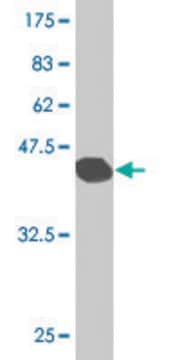
Evaluated by Western Blotting in rat lung tissue lysate.

Western Blotting Analysis: 1.0 µg/mL of this antibody detected SFTPC in 10 µg of rat lung tissue lysate.

Description de la cible

~20 kDa observed

Forme physique

Affinity purified
Purified rabbit polyclonal in buffer containing 0.1 M Tris-Glycine (pH 7.4), 150 mM NaCl with 0.05% sodium azide.

Stockage et stabilité

Stable for 1 year at 2-8°C from date of receipt.

Remarque sur l'analyse

Control
Rat lung tissue lysate

Autres remarques

Concentration: Please refer to the Certificate of Analysis for the lot-specific concentration.

Clause de non-responsabilité

Unless otherwise stated in our catalog or other company documentation accompanying the product(s), our products are intended for research use only and are not to be used for any other purpose, which includes but is not limited to, unauthorized commercial uses, in vitro diagnostic uses, ex vivo or in vivo therapeutic uses or any type of consumption or application to humans or animals.

Vous ne trouvez pas le bon produit ?  

Essayez notre Outil de sélection de produits.

Code de la classe de stockage

12 - Non Combustible Liquids

Classe de danger pour l'eau (WGK)

WGK 1

Point d'éclair (°F)

Not applicable

Point d'éclair (°C)

Not applicable


Certificats d'analyse (COA)

Recherchez un Certificats d'analyse (COA) en saisissant le numéro de lot du produit. Les numéros de lot figurent sur l'étiquette du produit après les mots "Lot" ou "Batch".

Déjà en possession de ce produit ?

Retrouvez la documentation relative aux produits que vous avez récemment achetés dans la Bibliothèque de documents.

Consulter la Bibliothèque de documents

William J Zacharias et al.
Nature, 555(7695), 251-255 (2018-03-01)
Functional tissue regeneration is required for the restoration of normal organ homeostasis after severe injury. Some organs, such as the intestine, harbour active stem cells throughout homeostasis and regeneration; more quiescent organs, such as the lung, often contain facultative progenitor
Shanru Li et al.
Stem cell reports, 18(9), 1841-1853 (2023-08-19)
AT2 cells harbor alveolar stem cell activity in the lung and can self-renew and differentiate into AT1 cells during homeostasis and after injury. To identify epigenetic pathways that control the AT2-AT1 regenerative response in the lung, we performed an organoid
Santiago Naranjo et al.
Genes & development, 36(15-16), 936-949 (2022-09-30)
Lung cancer is the leading cause of cancer-related death worldwide. Lung adenocarcinoma (LUAD), the most common histological subtype, accounts for 40% of all cases. While existing genetically engineered mouse models (GEMMs) recapitulate the histological progression and transcriptional evolution of human
Maria Plataki et al.
JCI insight, 5 (2019-07-10)
The prevalence of obesity is rising worldwide and obese patients comprise a specific population in the intensive care unit. Acute respiratory distress syndrome (ARDS) incidence is increased in obese patients. Exposure of rodents to hyperoxia mimics many of the features
Kuei-Pin Chung et al.
Nature communications, 15(1), 7241-7241 (2024-08-23)
Type 2 alveolar epithelial (AT2) cells of the lung are fundamental in regulating alveolar inflammation in response to injury. Impaired mitochondrial long-chain fatty acid β-oxidation (mtLCFAO) in AT2 cells is assumed to aggravate alveolar inflammation in acute lung injury (ALI)

Notre équipe de scientifiques dispose d'une expérience dans tous les secteurs de la recherche, notamment en sciences de la vie, science des matériaux, synthèse chimique, chromatographie, analyse et dans de nombreux autres domaines..

Contacter notre Service technique