Skip to Content
Merck
All Photos(5)

Documents

HPA012800

Sigma-Aldrich

Anti-PLD3 antibody produced in rabbit

enhanced validation

Prestige Antibodies® Powered by Atlas Antibodies, affinity isolated antibody, buffered aqueous glycerol solution

Synonym(s):

Anti-Choline phosphatase 3, Anti-HindIII K4L homolog, Anti-Hu-K4, Anti-PLD 3, Anti-Phosphatidylcholine-hydrolyzing phospholipase D3, Anti-Phospholipase D3

Sign Into View Organizational & Contract Pricing


About This Item

UNSPSC Code:
12352203
Human Protein Atlas Number:
NACRES:
NA.41

biological source

rabbit

conjugate

unconjugated

antibody form

affinity isolated antibody

antibody product type

primary antibodies

clone

polyclonal

product line

Prestige Antibodies® Powered by Atlas Antibodies

form

buffered aqueous glycerol solution

species reactivity

human

enhanced validation

orthogonal RNAseq
Learn more about Antibody Enhanced Validation

technique(s)

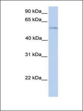
immunoblotting: 0.04-0.4 μg/mL
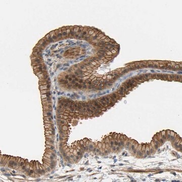
immunohistochemistry: 1:200-1:500

immunogen sequence

LDFPNASTGNPSTSQAWLGLLAGAHSSLDIASFYWTLTNNDTHTQEPSAQQGEEVLRQLQTLAPKGVNVRIAVSKPSGPQPQADLQALLQSGAQVRMVDMQKLTHGVLHTKFWVVDQTHFYLGSAN

UniProt accession no.

shipped in

wet ice

storage temp.

−20°C

target post-translational modification

unmodified

Gene Information

human ... PLD3(23646)

General description

Phospholipase D3 (PLD3) is a ubiquitously expressed type 2 transmembrane protein. It is present in the endoplasmic reticulum. The gene encoding this protein is located on chromosome 19q13.2.

Immunogen

Phospholipase D3 recombinant protein epitope signature tag (PrEST)

Application

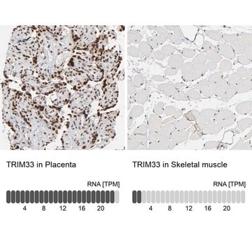
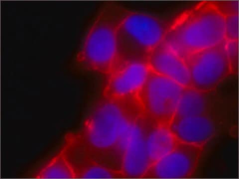
Anti-PLD3 antibody produced in rabbit, a Prestige Antibody, is developed and validated by the Human Protein Atlas (HPA) project . Each antibody is tested by immunohistochemistry against hundreds of normal and disease tissues. These images can be viewed on the Human Protein Atlas (HPA) site by clicking on the Image Gallery link. The antibodies are also tested using immunofluorescence and western blotting. To view these protocols and other useful information about Prestige Antibodies and the HPA, visit sigma.com/prestige.
Applications in which this antibody has been used successfully, and the associated peer-reviewed papers, are given below.
Western Blotting (1 paper)

Biochem/physiol Actions

Phospholipase D3 (PLD3) influences amyloid-β precursor protein (APP) processing. It is highly expressed in those regions of the brain which are vulnerable to Alzheimer′s disease pathology, including hippocampus and cortex. Overexpression of PLD3 leads to the decrease in intracellular APP and extracellular Aβ42 and Aβ40 (the isoforms of the amyloid-β peptide). On the other hand, knockdown of PLD3 leads to an increase in extracellular Aβ42 and Aβ40.

Features and Benefits

Prestige Antibodies® are highly characterized and extensively validated antibodies with the added benefit of all available characterization data for each target being accessible via the Human Protein Atlas portal linked just below the product name at the top of this page. The uniqueness and low cross-reactivity of the Prestige Antibodies® to other proteins are due to a thorough selection of antigen regions, affinity purification, and stringent selection. Prestige antigen controls are available for every corresponding Prestige Antibody and can be found in the linkage section.

Every Prestige Antibody is tested in the following ways:
  • IHC tissue array of 44 normal human tissues and 20 of the most common cancer type tissues.
  • Protein array of 364 human recombinant protein fragments.

Linkage

Corresponding Antigen APREST71731

Physical form

Solution in phosphate-buffered saline, pH 7.2, containing 40% glycerol and 0.02% sodium azide

Legal Information

Prestige Antibodies is a registered trademark of Merck KGaA, Darmstadt, Germany

Disclaimer

Unless otherwise stated in our catalog or other company documentation accompanying the product(s), our products are intended for research use only and are not to be used for any other purpose, which includes but is not limited to, unauthorized commercial uses, in vitro diagnostic uses, ex vivo or in vivo therapeutic uses or any type of consumption or application to humans or animals.

Not finding the right product?  

Try our Product Selector Tool.

Storage Class Code

10 - Combustible liquids

WGK

WGK 1

Personal Protective Equipment

dust mask type N95 (US), Eyeshields, Gloves

Certificates of Analysis (COA)

Search for Certificates of Analysis (COA) by entering the products Lot/Batch Number. Lot and Batch Numbers can be found on a product’s label following the words ‘Lot’ or ‘Batch’.

Already Own This Product?

Find documentation for the products that you have recently purchased in the Document Library.

Visit the Document Library

PLD3 gene and processing of APP.
Pietro Fazzari et al.
Nature, 541(7638), E1-E2 (2017-01-28)
Peng Yuan et al.
Nature, 612(7939), 328-337 (2022-12-01)
The precise mechanisms that lead to cognitive decline in Alzheimer's disease are unknown. Here we identify amyloid-plaque-associated axonal spheroids as prominent contributors to neural network dysfunction. Using intravital calcium and voltage imaging, we show that a mouse model of Alzheimer's
Yvette Roske et al.
Nucleic acids research, 52(1), 370-384 (2023-11-23)
The phospholipase D (PLD) family is comprised of enzymes bearing phospholipase activity towards lipids or endo- and exonuclease activity towards nucleic acids. PLD3 is synthesized as a type II transmembrane protein and proteolytically cleaved in lysosomes, yielding a soluble active
Jun-Ichi Satoh et al.
Alzheimer's research & therapy, 6(9), 70-70 (2014-12-06)
Recently, a whole-exome sequencing (WES) study showed that a rare variant rs145999145 composed of p.Val232Met located in exon 7 of the phospholipase D3 (PLD3) gene confers a doubled risk for late-onset Alzheimer's disease (AD). Knockdown of PLD3 elevates the levels
Thomas P Chapman et al.
Frontiers in immunology, 10, 1495-1495 (2019-08-06)
The interplay between NOD2 and TLR2 following recognition of components of the bacterial cell wall peptidoglycan is well-established, however their role in redirecting metabolic pathways in myeloid cells to degrade pathogens and mount antigen presentation remains unclear. We show NOD2

Our team of scientists has experience in all areas of research including Life Science, Material Science, Chemical Synthesis, Chromatography, Analytical and many others.

Contact Technical Service