FLAG® Tag Bacterial and Mammalian Expression Vectors
- FLAG® Tag Expression Vector Maps and 3xFLAG® Peptides
- FLAG® and 3xFLAG® Epitope Tag DNA Sequence and Amino Acid Peptide Sequence
- FLAG® and 3x FLAG® Bacterial and Mammalian Expression Vector Products
- FLAG® and 3xFLAG®: A Proven System for Detection and Purification of Proteins
- Highly Specific Antibodies to FLAG® and 3xFLAG® epitopes
- SnapFast™ FLAG® and 3xFLAG® Vector Legacy Product Comparison Chart
FLAG® Tag Expression Vector Maps and 3xFLAG® Peptides
Bacterial and mammalian expression vectors, or expression constructs, are circularized DNA transfection plasmids that are commonly used to overexpress recombinant proteins of interest. Unique sequences encoded within the vector construct contain promoter elements that are recognized by the transfected cells own protein synthesis machinery to drive the expression of the incorporated protein sequence.
The FLAG® Expression System is an established way to express, purify and detect recombinant fusion proteins. FLAG® and 3xFLAG® reagents have proven utility in numerous applications such as Western blotting, immunocytochemistry, immunoprecipitation, flow cytometry, protein purification, the study of protein-protein interactions, cell ultrastructure and protein localization. These small hydrophilic tags facilitate superior detection and purification of recombinant fusion proteins when using our highly specific and sensitive ANTI-FLAG® antibodies.
FLAG® and 3xFLAG® Epitope Tag DNA Sequence and Amino Acid Peptide Sequence
The FLAG® expression system utilizes a short, hydrophilic 8-amino acid peptide that is fused to the recombinant protein of interest. Because of its hydrophilic nature, the FLAG® peptide is likely to be located on the surface of the fusion protein. As a result, the FLAG® peptide is easily accessible for cleavage by enterokinase (Ek) and for detection with antibodies. In addition, because of the small size of the FLAG® peptide tag, it is not likely to obscure other epitopes, domains, or alter function, secretion, or transport of the fusion protein.
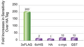
The 3xFLAG® system is an improvement upon the original system by fusing 3 tandem FLAG® epitopes (22 amino acids). Detection of fusion proteins containing 3xFLAG® is enhanced up to 200 times more than any other system. Like the original FLAG® tag, 3xFLAG® is hydrophilic, contains an Ek cleavage site, and is relatively small. Therefore, the risk of altering protein function, blocking other epitopes or decreasing solubility is minimized.

FLAG® and 3xFLAG® Amino Acid Sequences
Removal of N-terminal FLAG® (8 amino acids) and 3xFLAG® (22 amino acids) tags is possible using enterokinase, which cleaves following the Asp-Asp-Asp-Asp-Lys amino acid sequence at the c-terminal end of the tags.
FLAG® and 3x FLAG® Bacterial and Mammalian Expression Vector Products
The classic FLAG® and 3xFLAG® expression vectors have been converted to SnapFast™ Expression Vectors – combining two highly functional systems into one product. Additional information about the SnapFast™ system includes the Background & Popular Vectors, Selection Charts, and the SnapFast™ FAQs.
The FLAG® and 3xFLAG® vectors are available for bacterial and mammalian expression, using T7, Tac or CMV promoters. Vectors have an enterokinase cleavage site for removal of the tag from purified protein. Together with c-myc, some vectors offer dual-labeling. See charts below for quick-reference vector specifications and click the links to the product pages for more information, including vector maps.
FLAG® and 3xFLAG®: A Proven System for Detection and Purification of Proteins
Low-level expression of recombinant proteins in mammalian cells is a common problem that often makes detection of epitope tags difficult or impossible. Detection of 3x FLAG® using the ANTI-FLAG® M2 Ab and anti-mouse-HRP is 20-200 times more sensitive than any commonly used tag. An extensive selection of p3xFLAG® vectors utilizes the strong CMV promoter to drive expression.

Western blot of purified 3xFLAG® bacterial alkaline phosphatase and FLAG® bacterial alkaline phosphatase transferred on to nitrocellulose membrane. Detection was performed with ANTI-FLAG® M2 monoclonal antibody as primary, anti-mouse-HRP secondary antibody, and ECL™ chemiluminescent substrate.

C-terminal fusions of GST and the tags listed above were prepared and analyzed on Western Blots. All proteins were detected using the appropriate tag antibodies, anti-mouse HRP and ECL.
Highly Specific Antibodies to FLAG® and 3xFLAG® epitopes
The FLAG® and 3xFLAG® sequences include the binding sites for several highly specific ANTI-FLAG® monoclonal (M1, M2, M5) and polyclonal antibodies and conjugates, each with different recognition and binding characteristics. ANTI-FLAG® antibodies exhibit little or no cross-reactivity in most mammalian and bacterial cell lysates. One-step purification formats include affinity gels and 96-well plates.
SnapFast™ FLAG® and 3xFLAG® Vector Legacy Product Comparison Chart
Note: if you have used a FLAG® vector in the past, this chart will help you pick the closest SnapFast™ FLAG® and 3xFLAG® vector.
To continue reading please sign in or create an account.
Don't Have An Account?