Skip to Content
MilliporeSigma
All Photos(1)

Documents

G6642

Sigma-Aldrich

Anti-Glutamate antibody produced in rabbit

whole antiserum

Synonym(s):

Anti-Glutamate Ab, Glutamate Detection Antibody, Rabbit Anti-Glutamate

Sign Into View Organizational & Contract Pricing


About This Item

MDL number:
UNSPSC Code:
12352203
NACRES:
NA.41

biological source

rabbit

conjugate

unconjugated

antibody form

whole antiserum

antibody product type

primary antibodies

clone

polyclonal

contains

15 mM sodium azide

species reactivity

wide range

technique(s)

dot blot: 1:15,000

shipped in

dry ice

storage temp.

−20°C

target post-translational modification

unmodified

Related Categories

General description

The amino acids L-glutamate (Glu) and L-aspartate (Asp) are considered the major excitatory neurotransmitters in the central nervous system (CNS) and represent the most abundant mammalian neurotransmitter class. Both L-glutamate and L-aspartate are present in the brain at high concentrations and are distributed in most excitatory pathways in the CNS. Glu- and Asp-immunoreactivities are localized in high concentrations in synaptic terminals. In nerve terminals, L-glutamate is formed by deamidation of its major precursor, L-glutamine, by the enzyme glutaminase.
The actions of the excitatory amino acids on neurons are mediated by different receptor subtypes. These receptors are coupled to integral ion channels or to a second messenger system which utilizes inositol triphosphate (IP3). L-glutamate and L-aspartate may play an important role in the pathogenesis of certain neurological disorders such as Huntington′s disease, Alzheimer′s disease, epilepsy and brain ischemia. The excitoxic and neurotoxic effects of L-glutamate, leading to extensive neuronal damage, appear to be mediated by the N-methyl-D-aspartate (NMDA) receptor subtype.

Specificity

The antiserum is evaluated for activity and specificity by dot-blot immunoassay. The antiserum recognizes L-glutamic acid immobilized on an affinity membrane. No cross-reaction is observed with L-aspartic acid, L-glutamine, L-asparagine, and L-alanine. Weak cross-reactivity is observed with Gly-Asp, GABA, β-alanine, glycine and 5-aminovaleric acid (amino acid concentration 5-10 mM). Weak to moderate reaction is observed with L-glutamic acid-containing dipeptides Asp-Glu and Gly-Glu. The antibody may be used to localize glutamate in cell bodies, axons, and terminals of glutamatergic neurons in the CNS.

Immunogen

L-glutamic acid (Glu).

Application

Applications in which this antibody has been used successfully, and the associated peer-reviewed papers, are given below.
Immunohistochemistry (1 paper)
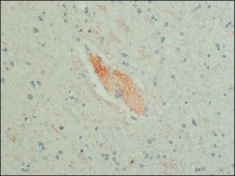
Rabbit polyclonal antiserum to L-glutamate may be used in immunohistochemical techniques on formalin- or glutaraldehyde-fixed, vibratome or frozen sections of human or animal tissues. Immunohistochemical methods provide increased anatomical resolution over conventional biochemical methods.

Disclaimer

Unless otherwise stated in our catalog or other company documentation accompanying the product(s), our products are intended for research use only and are not to be used for any other purpose, which includes but is not limited to, unauthorized commercial uses, in vitro diagnostic uses, ex vivo or in vivo therapeutic uses or any type of consumption or application to humans or animals.

Not finding the right product?  

Try our Product Selector Tool.

Storage Class Code

10 - Combustible liquids

WGK

WGK 3

Flash Point(F)

Not applicable

Flash Point(C)

Not applicable


Certificates of Analysis (COA)

Search for Certificates of Analysis (COA) by entering the products Lot/Batch Number. Lot and Batch Numbers can be found on a product’s label following the words ‘Lot’ or ‘Batch’.

Already Own This Product?

Find documentation for the products that you have recently purchased in the Document Library.

Visit the Document Library

Juan Guo et al.
EBioMedicine, 65, 103272-103272 (2021-03-11)
Despite the fundamental clinical significance of general anaesthesia, the cortical mechanism underlying anaesthetic-induced loss of consciousness (aLOC) remains elusive. Here, we measured the dynamics of two major cortical neurotransmitters, gamma-aminobutyric acid (GABA) and glutamate, through in vivo two-photon imaging and
Wenjun Zhou et al.
Nature neuroscience, 22(2), 205-217 (2019-01-22)
Nuclear receptor corepressor 1 (NCOR1) and NCOR2 (also known as SMRT) regulate gene expression by activating histone deacetylase 3 through their deacetylase activation domain (DAD). We show that mice with DAD knock-in mutations have memory deficits, reduced anxiety levels, and
Èlia Benito-Gutiérrez et al.
BMC biology, 19(1), 110-110 (2021-05-23)
The evolutionary origin of the telencephalon, the most anterior part of the vertebrate brain, remains obscure. Since no obvious counterpart to the telencephalon has yet been identified in invertebrate chordates, it is difficult to trace telencephalic origins. One way to
Marta Słoniecka et al.
PloS one, 10(7), e0134157-e0134157 (2015-07-28)
Keratocytes, the quiescent cells of the corneal stroma, play a crucial role in corneal wound healing. Neuropeptides and neurotransmitters are usually associated with neuronal signaling, but have recently been shown to be produced also by non-neuronal cells and to be
Pingwen Xu et al.
The Journal of clinical investigation, 125(7), 2861-2876 (2015-06-23)
Estrogen receptor-α (ERα) activity in the brain prevents obesity in both males and females. However, the ERα-expressing neural populations that regulate body weight remain to be fully elucidated. Here we showed that single-minded-1 (SIM1) neurons in the medial amygdala (MeA)

Our team of scientists has experience in all areas of research including Life Science, Material Science, Chemical Synthesis, Chromatography, Analytical and many others.

Contact Technical Service