Skip to Content
Merck
All Photos(1)

Key Documents

CBL1346Z-I

Sigma-Aldrich

Anti-mouse Integrin alphaV Antibody, clone RMV-7 (Preservative free)

clone RMV-7, from rat

Synonym(s):

Integrin alpha-V, Vitronectin receptor subunit alpha, CD51, 1.Integrin alpha-V heavy chain2.Integrin alpha-V light chain, Integrin alphaV

Sign Into View Organizational & Contract Pricing


About This Item

UNSPSC Code:
12352203
eCl@ss:
32160702
NACRES:
NA.41

biological source

rat

Quality Level

antibody form

purified immunoglobulin

antibody product type

primary antibodies

clone

RMV-7, monoclonal

species reactivity

mouse

technique(s)

flow cytometry: suitable
immunoprecipitation (IP): suitable
western blot: suitable

isotype

IgG1κ

NCBI accession no.

UniProt accession no.

shipped in

dry ice

target post-translational modification

unmodified

Gene Information

mouse ... Itgav(16410)

General description

Integrin alphaV, also known as CD51, a member of the integrin family, is a 140 kD protein expressed on activated T cells, polymorphonuclear granulocytes, blastocysts, and osteoclasts. Integrin alphaV forms heterodimers by association with integrins beta1, beta3, beta5 or beta6, these complexes then act as receptors for multiple extracellular matrix proteins (ECM). The alphaV integrins have varied functions in development, stimulation/activation and homeostasis. The primary ligands for Integrin alphaV complexes are fibronectin, fibrinogen, vitronectin, thrombspondin, von Willebrand factor, and CD31.

Specificity

Specifically recognizes Integrin alphaV, also known as CD51 or vitronectin receptor. This antibody has been reported to block binding of Integrin alphaV to vitronectin, fibronectin, fibrinogen, and CD31 in some cell types, as well as, blocking LAK cell cytotoxicity.

Immunogen

BALB/c mouse IL-2–activated killer (LAK) cells

Application

This Anti-mouse Integrin alphaV Antibody, clone RMV-7 (Preservative free) is validated for use in Western Blotting, Immunoprecipitation, Flow Cytometry for the detection of mouse Integrin alphaV.
Western Blot Analysis: A representative lot was used by an independent laboratory in LAK cell lysate. (Takahashi, K. et al., (1990). J. Immunol. 145:4371.)

Immunoprecipitation Analysis: A representative lot was used by an independent laboratory in LAK cell lysate. (Takahashi, K. et al., (1990). J. Immunol. 145:4371.)

Blocking Assay: Adhesion and LAK cell cytotoxicity was shown using a representative lot (Takahashi, K. et al., (1990). J. Immunol. 145:4371.)

Quality

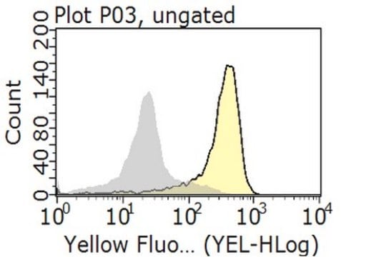
Evaluated by Flow Cytometry in murine 3T3-L1 cells.

Flow Cytometry Analysis: 0.2 µg of this antibody detected Integrin alphaV in murine 3T3-L1 cells .

Target description

~140 kDa observed. The calculated molecular weight is 115 kDa, however Integrin alphaV has been shown as a ~140 kDa band in western blots (Takahashi, K. et al., (1990). J. Immunol. 145:4371.).

Linkage

Replaces: CBL1346Z

Physical form

Format: Purified

Analysis Note

Control
Murine 3T3-L1 cells

Other Notes

Concentration: Please refer to the Certificate of Analysis for the lot-specific concentration.

Not finding the right product?  

Try our Product Selector Tool.

Storage Class Code

12 - Non Combustible Liquids

WGK

WGK 2

Flash Point(F)

Not applicable

Flash Point(C)

Not applicable


Certificates of Analysis (COA)

Search for Certificates of Analysis (COA) by entering the products Lot/Batch Number. Lot and Batch Numbers can be found on a product’s label following the words ‘Lot’ or ‘Batch’.

Already Own This Product?

Find documentation for the products that you have recently purchased in the Document Library.

Visit the Document Library

K Takahashi et al.
Journal of immunology (Baltimore, Md. : 1950), 145(12), 4371-4379 (1990-12-15)
CD2 and lymphocyte function-associated antigen (LFA)-1 are well known as T cell adhesion molecules involved in killer-target cell interactions. However, our recent study revealed that molecule(s) other than CD2 and LFA-1 might be involved in the lymphokine-activated killer (LAK) cell
Yongen Chang et al.
Journal of the American Society of Nephrology : JASN, 28(7), 1998-2005 (2017-02-22)
Activated fibroblasts are deemed the main executors of organ fibrosis. However, regulation of the pathologic functions of these cells in vivo is poorly understood. PDGF receptor β (PDGFRβ) is highly expressed in activated pericytes, a main source of fibroblasts. Studies

Our team of scientists has experience in all areas of research including Life Science, Material Science, Chemical Synthesis, Chromatography, Analytical and many others.

Contact Technical Service