Conducting Polymer Device Applications
Leslie H. Jimison1, Dion Khodagholy1, Thomas Doublet1,2, Christophe Bernard2, George G. Malliaras1, Róisín M. Owens1
1Department of Bioelectronics, Ecole Nationale Supérieure des Mines CMP-EMSE, MOC, 880 route de Mimet, 13541 Gardanne, France (www.bel.emse.fr), 2Institut de Neurosciences des Systèmes, Inserm UMR1106,Aix-Marseille Université Faculté de Médecine, 27 Boulevard Jean Moulin, 13005 Marseille, France
The application of conducting polymers at the interface with biology is an exciting new trend in organic electronics research 1-3 The nascent field of organic bioelectronics involves the coupling of organic electronic devices (such as electrodes and transistors) with biological systems, in an effort to bridge the biotic/abiotic interface. There are a number of unique characteristics of conducting polymers that make them wellsuited for integration with living systems:
- Mixed conduction of both electronic and ionic charge carriers is a major advantage for communication with biological systems, which rely heavily on ionic fluxes.
- Conducting polymers form ideal interfaces with electrolyte solutions that lack native oxides and dangling bonds.
- These van der Waals bonded solids have “soft” mechanical properties that better match those of the majority of biological tissues, leading to a lower mechanical mismatch in implanted devices.
- As in other applications for conducting polymers, solution processing of these materials facilitates easy fabrication and unique form factors.
Solution-processibility becomes especially important when designing flexible devices or inexpensive single-use sensors. These characteristics combine to provide a new toolbox for interfacing electronics with biology, and more importantly, solving problems related to biodiagnostics and treatment of biological dysfunction.
Herein we discuss two examples of conducting polymers at the interface with life sciences, showcasing the versatility and multi-functionality of these materials. The first is the development of conducting polymer electrodes that provide high-quality recordings of brain activity. The electrodes are fabricated on a highly conformable array, allowing for intimate contact with the surface of the brain. The second introduces an organic electronic cell-based sensor, where living tissue acts as the broad first line of defense against a range of toxins. This sensor has a direct application in diagnostics and understanding the mechanisms of drug delivery.
Poly(3,4-ethylenedioxythiophene):poly (styrenesulfonate) (PEDOT:PSS) in Bioelectronics
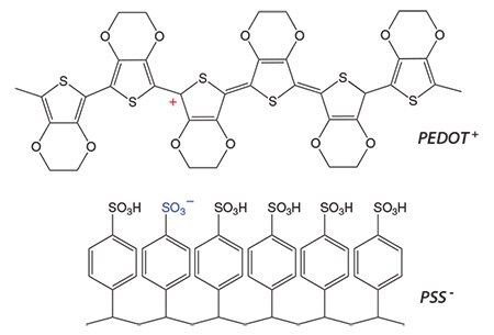
Due to the versatility of polymer synthesis, there exists a large library of conducting polymer systems. In recent years, PEDOT:PSS has emerged as a champion material in the field of bioelectronics. The p-conjugation gives PEDOT its semiconducting properties, while the PSS acts as a p-type dopant and raises the room temperature electrical conductivity to high levels (100 S/cm, even up to 1,000 S/cm with proper optimization). Both of the examples discussed in this article are based on commercially available PEDOT:PSS aqueous dispersions (Product Nos. 739332, 739324, 739316 and 768642).

Figure 1. Chemical structure of PEDOT+ and PSS-. The fixed negative charges on the PSS chain are balanced by polarons existing on the PEDOT chain, giving rise to enhanced electronic conductivity.
Applications in Brain Recordings
Electronic devices that interface with living tissue have become a necessity in clinics to improve diagnoses and treatments. One such example is electrocorticography (ECoG) electrode arrays, which are increasingly used for functional mapping of cognitive processes before certain types of brain surgery (e.g., tumors), for diagnostic purposes (e.g., epilepsy)5 and for brain-machine interfaces, an assistive technology for people with severe motor disabilities.6 These electrodes must conform to the curvilinear shapes of the brain and form high-quality electrical contacts. Thin sheets of polymeric materials with metal electrodes are traditionally used for this purpose.7
Using microfabrication techniques, our group prepared ECoG arrays with conducting polymer electrodes.8 A micrograph of the electrodes is shown in Figure 2A. The arrays consisted of a parylene substrate, gold contact pads and interconnects, and parylene insulation. A PEDOT:PSS film was deposited in appropriate holes in the insulation layer, defining the electrodes. The total thickness of the arrays was 4 μm, yet the electrode arrays had adequate mechanical strength to be selfsupporting and to be manipulated by a surgeon. Figure 2B shows a micrograph of an array conforming to the midrib of a small leaf.
Arrays were placed on the somatosensory cortex of anaesthetized rats. The recordings were done after the addition of bicuculline (Product No. 14340), a GABAA receptor antagonist that enables the genesis of sharp-wave events which mimic epileptic spikes.9 The recordings were validated against a commercial implantable electrode. Figure 2C shows that the power spectrum of a recording from a PEDOT:PSS electrode exhibits the typical 1/f property of the ECoG spectrum, with good definition of the 1–10 Hz and the 30 Hz bands. These bands are the dominant ones during bicuculline-triggered sharpwave events. It should be noted that gold electrodes of similar size did not yield adequate recordings, due to the fact that their interface impedance was much higher. Thus, PEDOT:PSS electrodes were found to outperform traditional metal electrodes, which highlights the importance of incorporating conducting polymers in a highly conformable electrode array format.

Figure 2. A) Micrograph of the array showing a detailed view of three electrodes. B) An electrode array is shown to conform to the midrib of a small leaf. C) Power spectrum of representative recording with a PEDOT:PSS electrode. The blue arrows indicate the 1–10 Hz and the 30 Hz bands.
Applications in Whole Cell Biosensors
We have demonstrated the direct integration of barrier tissue with an organic electrochemical transistor (OECT). The result is a cell-based sensor for monitoring barrier tissue integrity in situ with high sensitivity and temporal resolution.10 Barrier tissue is comprised of tightly packed layers of epithelial cells. Examples in the body include the gastrointestinal (GI) tract and the blood brain barrier. Acting as the first layer of defense, these cell layers help to block the passage of toxins and pathogens, but allow the passage of ions, water, and nutrients. The ability of barrier tissue to impose such highly regulated transport arises from the presence of protein complexes at the border between neighboring cells, including the tight junction (TJ) and the adherens junction (AJ).11 When these cell-cell seals are compromised, the regulated transport is affected as well. Thus, disruptions in barrier tissue integrity are often indicative of the presence of toxins and pathogens. The work presented here uses the Caco-2 cell line (Product No. 86010202), derived from a human colon cancer. When grown on a permeable membrane, these cells differentiate into an appropriate in vitro model for the gastrointestinal tract, characterized by localized junctional proteins, low permeability to tracer molecules, and high transepithelial electrical resistance (TER).12
The sensor presented here relies on the operating mechanism of an organic electrochemical transistor (OECT).13 An OECT consists of a conducting polymer (PEDOT:PSS) channel in contact with an electrolyte (here, cell culture medium). A gate electrode (Ag/AgCl) is immersed in the electrolyte, and source and drain contacts at either side of the transistor channel measure the drain current (ID). On application of a positive gate voltage (VG), cations from the electrolyte drift into the PEDOT:PSS channel, de-doping the conducting polymer and decreasing the drain current (ID). The ionic flux from the electrolyte into the polymer film determines the speed at which the drain current changes. By integrating the cell layer into the OECT architecture as shown in Figure 3A, the transient OECT behavior is directly correlated with the ionic flux allowed through the barrier tissue. A lower ionic flux results in a slower ID response and vice versa. Thus, changes in the barrier tissue integrity are reflected in changes in ID transient response.

Figure 3. A) Architecture of an OECT integrated with barrier tissue layer; tissue is supported on a permeable membrane. The illustration on the right shows tightly packed epithelial cells with junctional proteins that regulate transport. B) OECT characteristics, showing a VG pulse (top) and normalized drain current, ΔI/Io (bottom), when healthy cells are present (solid blue) and when no cells are present (dashed black). A reduction in ionic flux caused by the presence of a barrier tissue layer slows device response time (VG = 0.3 V). C) Normalized Response (NR) calculated from OECT modulation on addition of varying concentrations of ethylene glycol-bis(2-aminoethyl ether)-N,N,N’,N’-tetraacetic acid (EGTA), as shown. EGTA was added at time=0. NR=0 refers to observed modulation when a healthy barrier layer is present, and NR=1 refers to observed modulation when barrier properties are destroyed: progression from 0 to 1 indicates barrier tissue disruption.
We used the OECT barrier tissue sensor to monitor the effect of ethylene glycol-bis(2-aminoethylether)-N,N,N′,N′-tetraacetic acid (EGTA), a specific calcium chelator, on barrier tissue integrity (Product Nos. E3889 and 03777). The junctional protein complexes existing between cells are dynamic in nature: changes in the extracellular environment, including calcium concentration, affect their assembly and function. When the extracellular calcium levels are too low, some junctional proteins are internalized.14 EGTA has been shown previously to effectively modify extracellular environment, leading to an increase in paracellular diffusion across barrier tissue.15 On introduction of 1 mM EGTA to the basolateral side of the barrier tissue layer, we observe negligible changes in the response of the OECT, indicating no disruption of barrier properties. On addition of 10 and 100 mM EGTA, we observe a concentration-dependent effect, with the presence of 100 mM EGTA destroying barrier integrity within 45 minutes. With the OECT sensor, we are able to monitor the evolution of this disruption in 30-second increments. Results were validated with more traditional endpoint characterization methods including immunofluorescence staining of junctional proteins, permeability assays, and electrical impedance spectroscopy (data not shown). The sensitivity of the OECT sensor is equal to or greater than these established characterization techniques, and benefits from an extremely high temporal resolution. Moreover, the simple fabrication and device operation open up possibilities for creative solutions for large-scale diagnostics screening and in vitro model development.
Conclusions
In this article, we discussed two examples of novel organic bioelectronics devices: electrocorticography arrays and organic electrochemical transistors. Electrocorticography arrays designed with PEDOT:PSS electrodes outperform gold electrodes of similar geometry. These PEDOT:PSS based arrays provide a means of recording ECoG signals inside sulci in the human brain, significantly enhancing diagnostic capabilities, primarily due to their unique conformability. Organic electrochemical transistors deliver real-time information on subtle changes in barrier tissue integrity. The information obtained from this device can be used to understand the effect of pathogens and toxins, and the transport of drugs.
References
Pour continuer à lire, veuillez vous connecter à votre compte ou en créer un.
Vous n'avez pas de compte ?