Accéder au contenu
Merck
Toutes les photos(1)

Key Documents

MAB3414

Sigma-Aldrich

Anti-Cytokeratin 8 Antibody, clone 4.1.18

clone 4.1.18, Chemicon®, from mouse

Se connecterpour consulter vos tarifs contractuels et ceux de votre entreprise/organisme


About This Item

Code UNSPSC :
12352203
eCl@ss :
32160702
Nomenclature NACRES :
NA.41

Source biologique

mouse

Niveau de qualité

Forme d'anticorps

purified immunoglobulin

Type de produit anticorps

primary antibodies

Clone

4.1.18, monoclonal

Espèces réactives

human

Fabricant/nom de marque

Chemicon®

Technique(s)

immunocytochemistry: suitable
immunohistochemistry (formalin-fixed, paraffin-embedded sections): suitable
western blot: suitable

Isotype

IgG1

Numéro d'accès NCBI

Numéro d'accès UniProt

Conditions d'expédition

wet ice

Modification post-traductionnelle de la cible

unmodified

Informations sur le gène

human ... KRT8(3856)

Spécificité

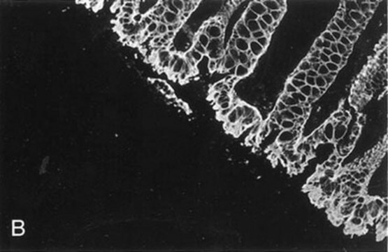
The antibody reacts with human cytokeratin 8. There is no cross reactivity with human cytokeratin No. 7, 17, 18, or 19. Anti-cytokeratin No. 8 decorates single epithelia in stomach, colon, and small intestine, gall bladder, liver (bile ducts and hepatocytes) as well as pancreatic ducts and mammary gland ducts.

Immunogène

Cytoskeletal preparation of MCF-7 cells.

Application

This Anti-Cytokeratin 8 Antibody, clone 4.1.18 is validated for use in WB, IC, IH, IH(P) for the detection of Cytokeratin 8.
Western blot Immunocytochemistry Immunohistochemistry: Frozen sections, acetone fixed (5-10 μg/mL)

Paraffin sections, after trypsin treatment (5-20 μg/mL)

Optimal-working dilutions must be determined by end user.

Liaison

Replaces: 04-588

Forme physique

Format: Purified
Lyophilized from 0.02 M Phosphate buffer, 0.25 M NaCl, pH 7.6 with 0.1% sodium azide.

Informations légales

CHEMICON is a registered trademark of Merck KGaA, Darmstadt, Germany

Vous ne trouvez pas le bon produit ?  

Essayez notre Outil de sélection de produits.

Code de la classe de stockage

10 - Combustible liquids

Classe de danger pour l'eau (WGK)

WGK 2

Point d'éclair (°F)

Not applicable

Point d'éclair (°C)

Not applicable


Certificats d'analyse (COA)

Recherchez un Certificats d'analyse (COA) en saisissant le numéro de lot du produit. Les numéros de lot figurent sur l'étiquette du produit après les mots "Lot" ou "Batch".

Déjà en possession de ce produit ?

Retrouvez la documentation relative aux produits que vous avez récemment achetés dans la Bibliothèque de documents.

Consulter la Bibliothèque de documents

Cristián Vela Hinojosa et al.
PloS one, 8(9), e75062-e75062 (2013-10-03)
The mammary gland has long drawn the attention of the scientific community due to the limited knowledge of some fundamental aspects involved in the control of its function. Myotis velifer, a microchiropteran species, provides an interesting model to study some
Stanislava Merjava et al.
Investigative ophthalmology & visual science, 52(2), 787-794 (2010-10-12)
The purpose of this study was to investigate the expression of cytokeratin (CK) 8 in the corneoconjunctival epithelium. In 17 cadaveric corneoscleral discs and 3 other discs, the presence of CK8 alone or CK8, together with CK3, CK15, vimentin, and
Miranda Batchelor et al.
EMBO reports, 5(1), 104-110 (2004-01-08)
While remaining extracellular, enteropathogenic Escherichia coli (EPEC) establish direct links with the cytoskeleton of the target epithelial cell leading to the formation of actin-rich pedestals underneath attached bacteria. The translocated adaptor protein Tir forms the transmembrane bridge between the cytoskeleton
Leandi Krüger et al.
American journal of physiology. Gastrointestinal and liver physiology, 313(3), G180-G191 (2017-06-03)
Esophageal injury is a risk factor for diseases such as Barrett's esophagus (BE) and esophageal adenocarcinoma. To improve understanding of signaling pathways associated with both normal and abnormal repair, animal models are needed. Traditional rodent models of esophageal repair are
Sinyoung Kim et al.
International journal of molecular medicine, 27(5), 701-706 (2011-02-25)
Although recent studies have demonstrated the in vitro hepatic differentiation potential of mesenchymal stem cells (MSCs), the evidence supporting the in vivo engraftment of MSCs, hepatic differentiation and improvement of hepatic function is still lacking. We investigated in vivo hepatic

Notre équipe de scientifiques dispose d'une expérience dans tous les secteurs de la recherche, notamment en sciences de la vie, science des matériaux, synthèse chimique, chromatographie, analyse et dans de nombreux autres domaines..

Contacter notre Service technique