Skip to Content
Merck
All Photos(3)

Documents

MABN504

Sigma-Aldrich

Anti-VDAC1 Antibody, clone N152B/23

clone N152B/23, from mouse

Synonym(s):

Voltage-dependent anion-selective channel protein 1, VDAC-1, hVDAC1, Outer mitochondrial membrane protein porin 1, Plasmalemmal porin, Porin 31HL, Porin 31HM

Sign Into View Organizational & Contract Pricing


About This Item

UNSPSC Code:
12352203
eCl@ss:
32160702
NACRES:
NA.41

biological source

mouse

Quality Level

antibody form

purified antibody

antibody product type

primary antibodies

clone

N152B/23, monoclonal

species reactivity

rat, mouse, human

packaging

antibody small pack of 25 μg

technique(s)

immunohistochemistry: suitable
western blot: suitable

isotype

IgG2aκ

NCBI accession no.

UniProt accession no.

shipped in

ambient

storage temp.

2-8°C

target post-translational modification

unmodified

Gene Information

human ... VDAC1(7416)

General description

Voltage-dependent anion-selective channel protein 1 (VDAC1, VDAC), also known as outer mitochondrial membrane protein porin 1, is the outer mitochondrial membrane receptor for hexokinase and BCL2L1. VDAC1 forms a channel through the mitochondrial membrane and is involved in small molecule diffusion, cell volume regulation and apoptosis. VDAC1 may participate in the formation of the permeability transition pore complex (PTPC), which is responsible for the release of mitochondrial products that triggers apoptosis.

Specificity

This antibody does not cross-react with VDAC2 or VDAC3 (Prof. J. Trimmer, University of California, Davis).

Immunogen

Recombinant protein corresponding to human VDAC1.

Application

Detect VDAC using this mouse monoclonal antibody, Anti-VDAC1 Antibody, clone N152B/23 validated for use in western blotting & IHC.
Research Category
Neuroscience
Research Sub Category
Developmental Signaling
Western Blotting Analysis: A representative lot states VDAC1 does not cross-react with VDAC2 or VDAC3 (based on KO validation results) representative lot deteted VDAC1 in wild type mouse liver tissue, and demonstrated a loss of signal in VDAC1 knock out mouse hippocampal tissue using immunobloting analysis (Prof. J. Trimmer, University of California, Davis).
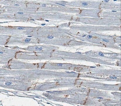
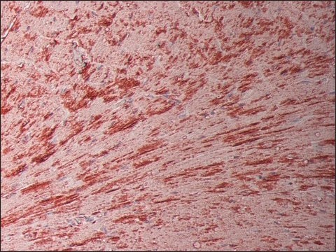
Immunohistochemistry Analysis: A 1:250 dilution from a representative lot detected VDAC1 in human cardiac myocytes tissue.

Quality

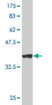
Evaluated by Western Blotting in mouse brain tissue lysate.

Western Blotting Analysis: 0.5 µg/mL of this antibody detected VDAC1 in 10 µg of mouse brain tissue lysate.

Target description

~ 35 kDa observed

Physical form

Format: Purified
Protein G Purified
Purified mouse monoclonal IgG2aκ in buffer containing 0.1 M Tris-Glycine (pH 7.4), 150 mM NaCl with 0.05% sodium azide.

Storage and Stability

Stable for 1 year at 2-8°C from date of receipt.

Analysis Note

Control
Mouse brain tissue lysate

Other Notes

Concentration: Please refer to the Certificate of Analysis for the lot-specific concentration.

Disclaimer

Unless otherwise stated in our catalog or other company documentation accompanying the product(s), our products are intended for research use only and are not to be used for any other purpose, which includes but is not limited to, unauthorized commercial uses, in vitro diagnostic uses, ex vivo or in vivo therapeutic uses or any type of consumption or application to humans or animals.

Not finding the right product?  

Try our Product Selector Tool.

Storage Class Code

12 - Non Combustible Liquids

WGK

WGK 1

Flash Point(F)

Not applicable

Flash Point(C)

Not applicable


Certificates of Analysis (COA)

Search for Certificates of Analysis (COA) by entering the products Lot/Batch Number. Lot and Batch Numbers can be found on a product’s label following the words ‘Lot’ or ‘Batch’.

Already Own This Product?

Find documentation for the products that you have recently purchased in the Document Library.

Visit the Document Library

Maria Feola et al.
Communications biology, 4(1), 517-517 (2021-05-05)
Erythropoiesis involves complex interrelated molecular signals influencing cell survival, differentiation, and enucleation. Diseases associated with ineffective erythropoiesis, such as β-thalassemias, exhibit erythroid expansion and defective enucleation. Clear mechanistic determinants of what make erythropoiesis effective are lacking. We previously demonstrated that
Ngonidzashe B Madungwe et al.
American journal of translational research, 12(7), 3412-3428 (2020-08-11)
MPV17 is an inner mitochondrial membrane protein whose mutation results in mitochondrial DNA (mtDNA) depletion diseases such as neurohepatopathy. MPV17 is expressed in several organs including the liver and kidneys. Here, we investigated its role and mechanism of action in
Mitofilin Heterozygote Mice Display an Increase in Myocardial Injury and Inflammation after Ischemia/Reperfusion.
Feng, et al.
Antioxidants (Basel, Switzerland), 12 (2023)
E D'Acunto et al.
Cellular and molecular life sciences : CMLS, 79(8), 437-437 (2022-07-22)
The neurodegenerative condition FENIB (familiar encephalopathy with neuroserpin inclusion bodies) is caused by heterozygous expression of polymerogenic mutant neuroserpin (NS), with polymer deposition within the endoplasmic reticulum (ER) of neurons. We generated transgenic neural progenitor cells (NPCs) from mouse fetal
Arnaud Mourier et al.
The Journal of cell biology, 208(4), 429-442 (2015-02-18)
Mitochondria form a dynamic network within the cell as a result of balanced fusion and fission. Despite the established role of mitofusins (MFN1 and MFN2) in mitochondrial fusion, only MFN2 has been associated with metabolic and neurodegenerative diseases, which suggests

Our team of scientists has experience in all areas of research including Life Science, Material Science, Chemical Synthesis, Chromatography, Analytical and many others.

Contact Technical Service