Skip to Content
Merck
HomeWestern BlottingCell Lysate Preparation

Cell Lysate Preparation

Often, the first step of analyzing protein expression or protein-protein interactions is to obtain a cell or tissue sample, lyse the cells, and extract proteins using extraction reagents. Total protein concentration must be determined for these cell lysates. Variables affecting each of these steps are outlined below, as each could affect the sensitivity and reproducibility of the Western blot.

Click on the symptoms to read about the possible causes and remedies:

Total Protein Concentration Too Low

Protein concentration too low as seen on Coomassie-stained membrane

Protein concentration
too low as
seen on
Coomassie-stained
membrane
Possible CauseRemedy

Heat degradation
  • Perform all lysate preparation steps at 4 °C.
  • If sonicating, minimize total sonication time or insert breaks in between sonication pulses to minimize heat generation.

Protein degradation
  • Add or increase concentration of protease and phosphatase inhibitors.
  • Try using different inhibitor cocktails.
  • Create lysate aliquots to minimize number of freeze-thaw cycles.

Inadequate tissue homogenization
 
  • Increase time/power setting of sonication.

Cell debris contamination
  • When pipetting supernatant following centrifugation, take care not to disturb the pellet.

Too little tissue/too few cells
  • Use more tissue and/or less buffer.
  • If using dissociated/cultured cells, make sure cell counts are accurate.

Wrong type of lysis buffer

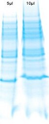
Effect of excessively low sample pH on SDS-PAGE

Different proteins may be best visualized via Western blot using different lysis buffers. Consider the pH, buffering compound, detergents and other additives when determining the optimal buffer for a specific 

Protein Concentration Too High

Possible Cause Remedy

Insufficient buffer/tissue ratio
  • Use less tissue and/or more buffer.
  • If using dissociated, cultured cells, make sure cell counts are accurate.

Variability in Measured Total Protein Concentration between Replicates

Possible Cause      Remedy

Inaccurate pipetting
  • Ensure accurate pipetting; use properly calibrated pipettes.

Incorrect protein standard selected (for colorimetric assays)
  • Switch to infrared-based protein quantitation, which directly measures amide bonds in protein chains

Time before reading absorbance too long (for colorimetric assays)
  • Process fewer samples at once.
  • Switch to infrared-based protein quantitation, which is not time-dependent.

Assay incompatible with buffers used
  • Verify that buffers used are compatible with chosen protein quantitation assay.
  • Use infrared-based protein quantitation, which is compatible with a wider range of buffers than colorimetric assays.

Protein concentration outside of linear dynamic range of assay
  • Increase or decrease lysate concentration until it is within the linear dynamic range of the selected assay.
  • Choose an assay with a broader linear dynamic range.

Non-Linear Standard Curve (for Colorimetric Assays for Total Protein)

Possible Cause                             Remedy

Pipetting error resulting in incorrect
dilution of standards.
  • Ensure accurate pipetting; use properly-calibrated pipettes.

Degradation Band on Blot

Possible CauseRemedy

Protein degradation due to insufficient
protease inhibition
  • Add or increase concentration of protease and phosphatase inhibitors.
  • Try using different inhibitor cocktails.

Bands Smeared Vertically

Possible CauseRemedy

Protein degradation
  • Increase concentration of protease and/or phosphatase inhibitors during cell lysate preparation.
  • Avoid repeated freeze/thaw cycles of cell lysate and Western blot samples.

Incompatibility between lysis
buffer component and SDS

Effect of interfering detergents (right lane) in SDS-PAGE

Effect of interfering detergents
(right lane) in SDS-PAGE
  • Cationic detergents in the lysis buffer will modify charge of SDS micelles, causing smearing. Remove or substitute these detergents.
  • Nonionic detergents at high concentrations will modify SDS micelles, causing smearing. Dilute, or remove these detergents.

Excess concentration of abundant proteins, such as albumin, in the sample
  • First deplete the protein sample of abundant proteins, using magnetic or agarose beads designed for depletion and/or enrichment

Bands of Interest Obscured

Bands of interest on SDS-PAGE obscured by abundant proteins

Bands of interest
on SDS-PAGE
obscured by
abundant proteins
Possible CauseRemedy

Excess concentration of abundant proteins, such as albumin, in the sample
  • First deplete the protein sample of abundant proteins, using magnetic or agarose beads designed for depletion and/or enrichment.

Materials
Sorry, an unexpected error has occurred

Network error: Failed to fetch

Sign In To Continue

To continue reading please sign in or create an account.

Don't Have An Account?