Journal of muscle research and cell motility, 5(1), 97-112 (1984-02-01)
The properties of spin-labelled myosin, prepared from rabbit skeletal and scallop adductor muscle, on forming a long-lived complex with ADP and vanadate (M.ADP.Vi), have been investigated. In the case of an iodoacetamide-based label attached to rabbit myosin or subfragment 1
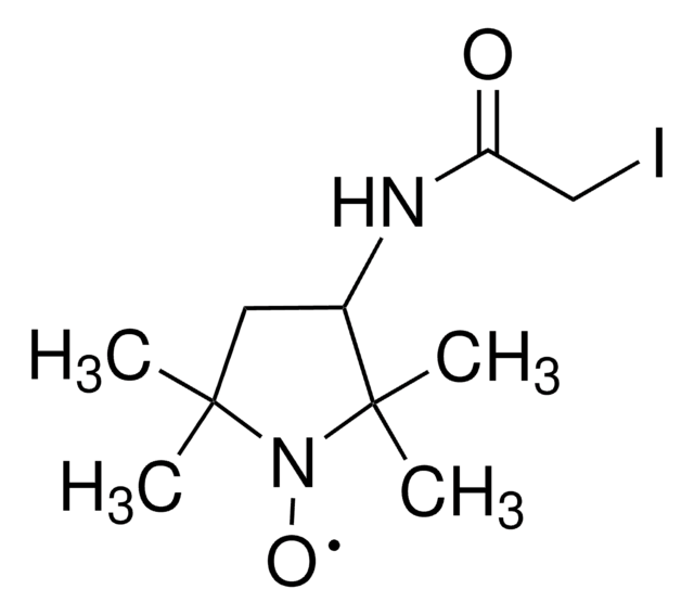
The labeling kinetics of sarcoplasmic reticulum ATPase with the iodoacetamide spin probe N-(1-oxy-2,2,6,6-tetramethyl-4-piperidinyl)iodoacetamide were followed under conditions designed to selectively label all reactive groups. Approximately 1 mol of spin-label reacted per one 100 000-dalton ATPase chain, indicating only one residue
British journal of industrial medicine, 41(1), 46-50 (1984-02-01)
Alterations in erythrocyte membranes caused by UICC B chrysotile asbestos fibres were studied in red cell ghosts using the spin label technique. The electron paramagnetic resonance (EPR) spectra of two sulphydryl reactive spin labels and one fatty acid spin probe
We have observed the effects of MgADP and thiophosphorylation on the conformational state of the light chain domain of myosin in skinned smooth muscle. Electron paramagnetic resonance (EPR) spectroscopy was used to monitor the orientation of spin probes attached to
Sarcoplasmic reticulum vesicles were labeled with [14C]iodoacetamide spin-label (ISL) under conditions where time courses of the reaction predicted that one amino acid residue would be preferentially labeled. Solubilized tryptic peptides were separated by high-performance liquid chromatography following extensive digestion, and
Our team of scientists has experience in all areas of research including Life Science, Material Science, Chemical Synthesis, Chromatography, Analytical and many others.